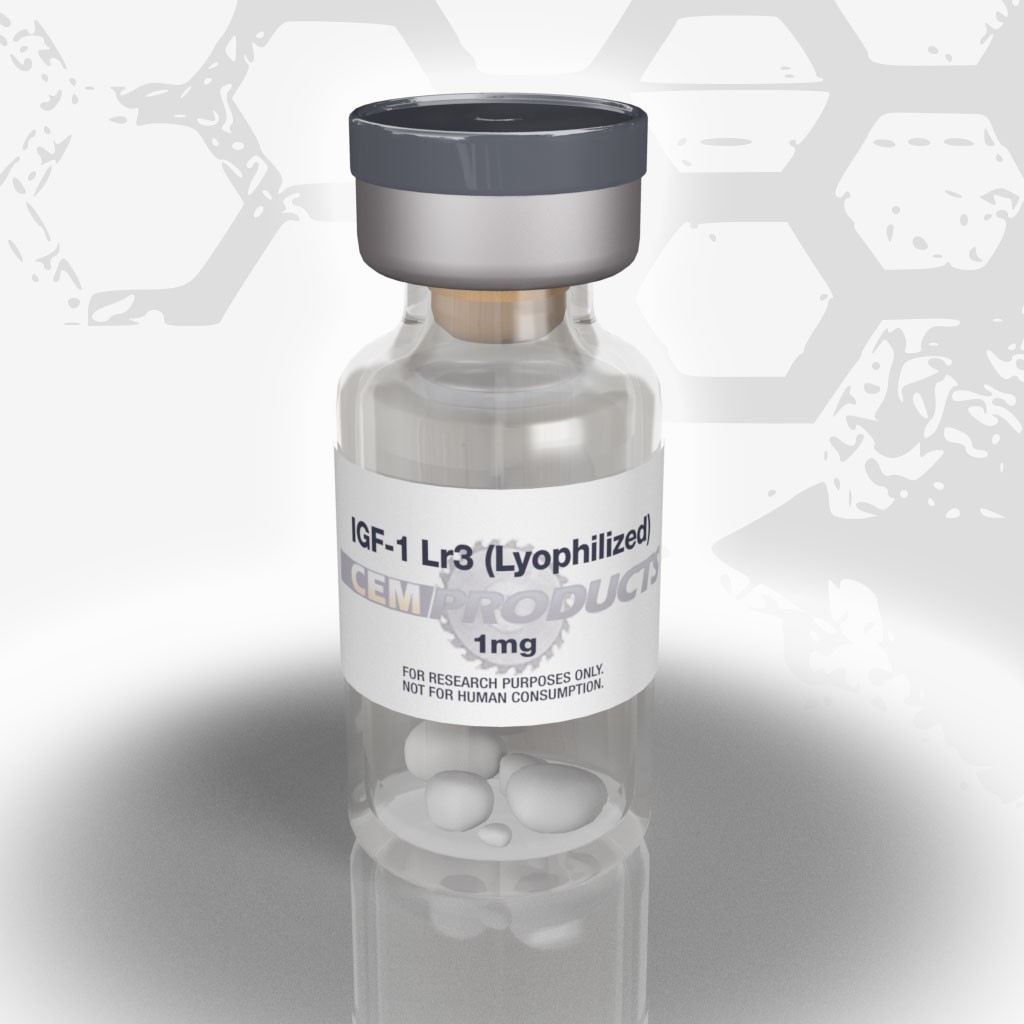
Localized infusion of IGF-I results in skeletal muscle hypertrophy in rats.
Researchers:Gregory R. Adams & Samuel A. McCue
Department of Physiology and Biophysics, University of California, Irvine, Ca.
Source:Journal of Applied Physiology84(5): 1716-1722, 1998
Summary:The present study was undertaken to test the hypothesis that direct IGF-I infusion would result in an increase in muscle DNA as well as in various measurements of muscle size. Either 0.9% saline or nonsystemic doses of recombinant human IGF-I (rhIGF-1) were infused directly into a non-weight-bearing muscle of rats, the tibialis anterior (TA), via a fenestrated catheter attached to a subcutaneous miniosmotic pump. Saline infusion had no effect on the mass, protein content, or DNA content of TA muscles. Local IGF-I infusion had no effect on body or heart weight. The absolute weight of the infused TA muscles was ~9% greater (P < 0.05) than that of the contra-lateral TA muscles. IGF-I infusion resulted in significant increases in the total protein and DNA content of TA muscles (P < 0.05). As a result of these coordinated changes, the DNA-to-protein ratio of the hypertrophied TA was similar to that of the contra-lateral muscles. These results suggest that IGF-I may be acting to directly stimulate processes such as protein synthesis and satellite cell proliferation, which result in skeletal muscle hypertrophy.
Discussion:The details of the mechanisms and pathways by which mechanical stress stimulates localized muscle fiber hypertrophy are still being elucidated. It is clear however, that growth hormone (GH), fibroblast growth factors (FGF) and insulin-like growth factors (IGF) play a central role in this process. Insulin-like growth factor I (IGF-I) peptide levels have been shown to increase in overloaded skeletal muscles (G. R. Adams and F. Haddad. J. Appl. Physiol. 81: 2509-2516, 1996). In that study, there was an increase in IGF-1 content before measurable increases in muscle protein and was correlated with an increase in muscle DNA content. Several other studies have shown that muscle fibers undergoing hypertrophy, due to mechanical stress, express elevated levels of IGF-I prior to hypertrophy.
IGF-1 appears to be an important regulator of the nuclear to cytoplasmic ratio. Studies have show that a muscle will only undergo hypertrophy if it can maintain the ratio of the cell’s volume to the number of nuclei within a finite limit. In the study above, a relatively “unloaded” muscle, the anterior tibialis, was injection with 0.9 – 1.9micrograms/kg/day of rhIGF-1 which then mimicked the effects of physically loading the muscle. There was an increase in protein content, cross sectional area and DNA content. The increase in muscle DNA is presumed to be a result of increased proliferation and differentiation of satellite cells which donate their nuclei upon fusion with damaged or hypertrophying muscle cells. Take note that the quantities of IGF-1 used in the injections were extremely small, much smaller than studies that have shown relatively poor results from administering IGF-1 systemically which range from 1.0 to 6.9milligrams/kg/day.
All of the attention and discussion of half-hazzardly injecting fat into muscles to increase the girth of a limb is only a symptom of the obsessive nature of bodybuilding. I would imagine that locally injecting minute amounts (micrograms) of rhIGF-1 to actually increase the growth of individual muscles would be a far better alternative to injecting fat, Esiclene or even getting silicone implants. Those bodybuilders at the national or professional level with lagging calves would be wise to consider the results of this study should they stumble across a bottle of Genentech’s rhIGF-1!

Leave a Reply
You must be logged in to post a comment.