Myostatin

OhNoYo

New Member
When Will We Have a SAFE & EFFECTIVE Myostatin Inhibitor in the Future?
Anyone??? :confused: :popcorn:
 
Last edited by a moderator:
Re: When Will We Have a SAFE & EFFECTIVE Myostatin Inhibitor in the Future?

if they ever make one that is real and works for real - steriods will no longer have a place in bodybuilding guys!

it is the truth - why mess with ur hormones when you could just alter ur body to create more muscle cells - haha but keep wishing nothing is real so far good at all that i know of - there is that dog and a couple animals and people with the gene issue

- there is 1 german boy that has double muscle and has the myostin gene problem - i wish i could find him online but i can find no new pix other than him as a baby - he COULD BE THE BEST BOSYBUILDER EVER NO JOKE THINK ABOUT IT

I disagree. To most, bodybuilding is about pushing the abnormal envelope. So if steroids are good...and an effective (and attainable) myostatin inhibitor is good...then 1+1=3, right? 400lb behemoth bodybuilders here we come!

Doesn't Flex Wheeler claim to have a myostatin defect? :D
 
Last edited:
Re: When Will We Have a SAFE & EFFECTIVE Myostatin Inhibitor in the Future?

I was surprised to not see more posts on this thread. After all, this is an area of active R&D and undoubtedly leads to increased musculature. Is it possible that many do not recall one the images first published (below)? This image is from the 1997 PNAS article, "McPherron AC, Lee S-J. Double muscling in cattle due to mutations in the myostatin gene. Proceedings of the National Academy of Sciences of the United States of America 1997;94(23):12457-61." http://www.pnas.org/content/94/23/12457.full.pdf

The original publication was in the same year that included the above authors, "McPherron AC, Lawler AM, Lee SJ. Regulation of skeletal muscle mass in mice by a new TGF-beta superfamily member. Nature 1997;387(6628):83-90." Regulation of skeletal muscle mass in mice by a new TGF-p superfamily member In this publication, we were introduced to the "Mighty Mouse."

Myostatin, a member of the transforming growth factor (TGF)-? superfamily, plays a potent inhibitory role in regulating skeletal muscle mass. Increasing size and strength of skeletal muscle represents a promising therapeutic strategy for muscular disorders. Inhibition of myostatin by gene disruption, transgenic expression of myostatin propeptide, or injection of propeptide or myostatin antibodies, causes a widespread increase in skeletal muscle mass. Several peptides, in addition to myostatin propeptide and myostatin antibodies, can bind directly to and neutralize the activity of myostatin. [More on this later.]

F2.medium.gif
 
Re: When Will We Have a SAFE & EFFECTIVE Myostatin Inhibitor in the Future?

I was surprised to not see more posts on this thread. After all, this is an area of active R&D and undoubtedly leads to increased musculature. Is it possible that many do not recall one the images first published (below)? This image is from the 1997 PNAS article, "McPherron AC, Lee S-J. Double muscling in cattle due to mutations in the myostatin gene. Proceedings of the National Academy of Sciences of the United States of America 1997;94(23):12457-61." http://www.pnas.org/content/94/23/12457.full.pdf

The original publication was in the same year that included the above authors, "McPherron AC, Lawler AM, Lee SJ. Regulation of skeletal muscle mass in mice by a new TGF-beta superfamily member. Nature 1997;387(6628):83-90." Regulation of skeletal muscle mass in mice by a new TGF-p superfamily member In this publication, we were introduced to the "Mighty Mouse."

Myostatin, a member of the transforming growth factor (TGF)-? superfamily, plays a potent inhibitory role in regulating skeletal muscle mass. Increasing size and strength of skeletal muscle represents a promising therapeutic strategy for muscular disorders. Inhibition of myostatin by gene disruption, transgenic expression of myostatin propeptide, or injection of propeptide or myostatin antibodies, causes a widespread increase in skeletal muscle mass. Several peptides, in addition to myostatin propeptide and myostatin antibodies, can bind directly to and neutralize the activity of myostatin. [More on this later.]

8766

great contribution to this thread, Doc. as for number of posts, i think many people r still in a 'wait n see' mode re: myostatin inhibition and genetic manipulation in general. the thread is obviously conjectural...many of us aren't comfortable conjecturing usefully on this question. :D

i would have to imagine there's some degree of unlicensed and unsupervised research going on...it's a pity when some good ideas dont get shared.
 
Re: When Will We Have a SAFE & EFFECTIVE Myostatin Inhibitor in the Future?

Here is the dog

so you can see steriods would not matter much at all - this dog does not take riod or even bodybuild and is pure muscle - so imagine if someone just ate clean and bodybuilded clean with this condition - they would be 300 lbs more ripped than jay cutler for real \

and here is the german child - he has muscle legs at 7 moths guys - think if he bodybuilds at 20 years old!!!!!
 

Attachments

  • post-7-57588-MyostatinDog2.jpg
    post-7-57588-MyostatinDog2.jpg
    29.4 KB · Views: 306
  • KID.jpg
    KID.jpg
    5 KB · Views: 329
  • KIDS.jpg
    KIDS.jpg
    4.3 KB · Views: 327
Re: When Will We Have a SAFE & EFFECTIVE Myostatin Inhibitor in the Future?

i disagree. To most, bodybuilding is about pushing the abnormal envelope. So if steroids are good...and an effective (and attainable) myostatin inhibitor is good...then 1+1=3, right? 400lb behemoth bodybuilders here we come!

Doesn't flex wheeler claim to have a myostatin defect? :d
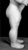
yes flex wheeler claims this or has in the past - but its pure bullsh*t

no tests have ever come to show he has the condition and if he did he would have no problems winning olympias - he would have been unbeatable with riods and this condition


no he doesnt but has claimed to in the past as a coverup for massive amounts of gear
 
Re: When Will We Have a SAFE & EFFECTIVE Myostatin Inhibitor in the Future?

Here is the dog

so you can see steriods would not matter much at all - this dog does not take riod or even bodybuild and is pure muscle - so imagine if someone just ate clean and bodybuilded clean with this condition - they would be 300 lbs more ripped than jay cutler for real \

and here is the german child - he has muscle legs at 7 moths guys - think if he bodybuilds at 20 years old!!!!!


A healthy woman who was a former professional athlete gave birth to a son after a normal pregnancy. The identity of the child's father was not revealed. The child's birth weight was in the 75th percentile. He appeared extraordinarily muscular, with protruding muscles in his thighs and upper arms. With the exception of increased tendon reflexes, the physical examination was normal. Hypoglycemia and increased levels of testosterone and insulin-like growth factor I were excluded. Muscular hypertrophy was verified by ultrasonography when the infant was six days of age. The stimulus-induced myoclonus gradually subsided after two months. The child's motor and mental development has been normal. Now, at 4.5 years of age, he continues to have increased muscle bulk and strength, and he is able to hold two 3-kg dumbbells in horizontal suspension with his arms extended.


Schuelke M, Wagner KR, Stolz LE, et al. Myostatin mutation associated with gross muscle hypertrophy in a child. N Engl J Med 2004;350(26):2682-8. http://www.nejm.org/doi/full/10.1056/NEJMoa040933#t=article

McNally EM. Powerful genes--myostatin regulation of human muscle mass. N Engl J Med 2004;350(26):2642-4. http://www.nejm.org/doi/full/10.1056/NEJMp048124#t=article
 

Attachments

Last edited:
Re: When Will We Have a SAFE & EFFECTIVE Myostatin Inhibitor in the Future?

How myostatin inhibition could be incorporated into the doping toolkit is unknown, but there are concerns that genetic manipulation of myostatin gene expression may be in the first wave of ‘‘gene doping.’’ Currently, the potential effects of anti-myostatin treatments on athletic performance are unclear, but a growing body of data has shown that commensurate increases in muscle strength and power may not accompany the cellular and histological changes resulting from inhibited myostatin function. This review highlights the function of myostatin in muscle growth, developments in myostatin-targeted pharmacological and gene therapies, and the potential of these therapies to be appropriated by unscrupulous athletes and their entourages looking for a short cut to excellence.


Fedoruk MN, Rupert JL. Myostatin inhibition: a potential performance enhancement strategy? Scandinavian Journal of Medicine & Science in Sports 2008;18(2):123-31. Myostatin inhibition: a potential performance enha... [Scand J Med Sci Sports. 2008] - PubMed result

A decade has passed since myostatin was first identified as a negative regulator of muscle growth. Since then, studies in both humans and animals have demonstrated that decreasing the levels of this growth factor or inhibiting its function can dramatically increase muscle size, and a number of therapeutic applications of myostatin inhibition to the treatment of myopathies and muscle atrophy have been proposed. As such treatments would be likely to also stimulate muscle growth in healthy individuals, there is a growing concern among anti-doping authorities that myostatin inhibitors may be among the next generation of ergogenic pharmaceuticals or even in the vanguard of “gene doping” technology. While the ability to stimulate muscle growth through myostatin inhibition is well documented, a growing body of evidence suggests such increases may not translate into an improvement in athletic performance. This article briefly reviews the function of this potent regulator of muscle development and explores the potential therapeutic uses, and potential ergogenic abuses, of myostatin manipulation.

:rolleyes:

"steroids don't work"

"myostatin-inhibition doesn't work"

"just say no"

[:o)]

:popcorn:
 
Re: When Will We Have a SAFE & EFFECTIVE Myostatin Inhibitor in the Future?

Doesn't the peptide follistatin ( sp?) have an effect on myostatin ?

Have a buddy who has gone through a few cycles of it, noted soreness in the injection spot and improved recovery. I'm too chicken to try something that new ( and I have cancer in my family)

Yes, follistatin is supposed to be a myostatin inhibitor.

Did your friend gain any noticeable lean muscle mass from using it though, or just improved recovery, Sir?
 
Re: When Will We Have a SAFE & EFFECTIVE Myostatin Inhibitor in the Future?

A big issue w/ myostatin inhibitors is understanding the full scope of the what they do. As w/ any pharma solution for something they come up w/ - you can't pick and choose your results & sides. The body works on a complex system of biochemical "push / pull" paths and it is very hard to isolate a particular result w/o getting into the peripheral dependencies.
 
[Myostatin] A Cohort Study Of Racing Performance In Thoroughbred Racehorses

Can anyone doubt that the very same associations will be found in humans. Further, will these traits be used [by countries] to screen for athleticism.


Thoroughbred horses originated from a small number of Arab, Barb and Turk stallions and native British mares ?300 years ago. During the past 300 years, Thoroughbred horses have been selectively bred to improve speed and stamina, making them superior competitive racehorses. Thoroughbred horses have a very high skeletal muscle mass comprising over 55% of their total body mass. The aerobic capacity of Thoroughbred horses (VO2max > 200 ml O2/kg/min) is superior to that of other athletic species of similar size. Such traits have been enhanced by intense artificial selection for sequence variants contributing to exceptional racing performance. Various measures for the evaluation of racing performance in Thoroughbred horses, such as earnings, race times and handicap ratings, have been used to estimate heritabilities, and breeding values have also been calculated to evaluate horses’ potential ability to transmit genetic factors related to racing performance. While most of these studies have estimated the genetic contribution to variation in racing ability to be between 0.35 and 0.55, the heritability of best race distance (BRD) has been estimated to be as high as 0.94.

Many significant advances have been achieved in the horse genome project, such as the construction of half- and full-sibling linkage maps, horse-human comparative maps, and the completion of a high-quality draft horse genome sequence with over 1.1 million identified Single Nucleotide Polymorphisms (SNPs). This genetic infrastructure for the horse has enabled the identification of a genomic region on ECA18 as being associated with racing performance phenotypes.

Four independent studies, including a candidate gene study, a microsatellite-based genome-wide association study, and two genome-wide SNP association studies, have identified the same genomic region on ECA18 associated with racing performance. In this region, four SNPs (g.65809482T>C, g.65868604G>T, g.66493737C>T and g.66539967A>G) were identified as candidates for genetic prediction of racing performance in Japanese racehorses based on lifetime earnings and performance rank.

In particular, the SNP g.66493737C>T, which is located in the first intron of the myostatin (MSTN) gene, has been associated with BRD among a cohort of elite race winning Thoroughbreds. The MSTN gene is known to contribute to muscle hypertrophy phenotypes in a variety of mammalian species. It was observed that Thoroughbreds with the C/C genotype at g.66493737C>T were better suited to short-distance races, C/T horses competed favourably in middle-distance races, and T/T horses had greater stamina and represented the majority of winning horses at the longer distances.

In this study, researchers designed a retrospective cohort study to evaluate the influences of the four SNPs associated with racing performance in Japanese Thoroughbred racehorses. The Japan Racing Association (JRA) is the largest racing authority in Japan and hosts horse races for ?50% (?4000 horses) of the Thoroughbred horse population born in Japan every year. In this study, 1710 Thoroughbred racehorses born in the same year (2000) were followed over their athletic career in JRA horse races to evaluate racing performance based on lifetime earnings, JRA performance rank, BRD, and win-race distance (WRD), defined as the average distance of races won. The findings of the current study corroborate those previously reported.

In summary, of the four SNPs, g.65809482T>C, g.65868604G>T and g.66493737C>T were found to have similar diagnostic effects for optimum race distance. This finding is supported by the strong linkage disequilibrium (r2 > 0.8) associating these three SNPs. In addition, the use of multiple SNPs, such as g.65809482T>C–g.66493737C>T and g.65868604G>T–g.66493737C>T, may lead to a more accurate genetic prediction of WRD and BRD. This finding suggests that a cis-regulatory element or an actual susceptible SNP for racing performance is located in the candidate genomic region on ECA18 at the MSTN gene locus.


Tozaki T, Hill EW, Hirota K, et al. A cohort study of racing performance in Japanese Thoroughbred racehorses using genome information on ECA18. Anim Genet 2012;43(1):42-52. A cohort study of racing performance in Japanese Thoroughbred racehorses using genome information on ECA18 - Tozaki - 2011 - Animal Genetics - Wiley Online Library

Using 1710 Thoroughbred racehorses in Japan, a cohort study was performed to evaluate the influence of genotypes at four single nucleotide polymorphisms (SNPs) on equine chromosome 18 (ECA18), which were associated in a previous genome-wide association study for racing performance with lifetime earnings and performance rank. In males, both g.65809482T>C and g.65868604G>T were related to performance rank (P = 0.005). In females, g.65809482T>C (P = 1.76E-6), g.65868604G>T (P = 6.81E-6) and g.66493737C>T (P = 4.42E-5) were strongly related to performance rank and also to lifetime earnings (P < 0.05). When win-race distance (WRD) among all winning racehorses and best race distance (BRD) among elite racehorses were considered as the phenotypes, significant associations (P < 0.001) were observed for all four SNPs. The favourable race distance of both elite (BRD) and novice racehorses (WRD) was also associated with genotypes in the ECA18 region, indicating the presence of a gene in this region influencing optimum race distance in Thoroughbred racehorses. Therefore, the association with performance rank is likely due to the bias in the race distances. The location of the SNPs within and proximal to the gene encoding myostatin (MSTN) strongly suggests that regulation of the MSTN gene affects racing performance. In particular, the g.65809482T>C, g.65868604G>T and g.66493737C>T SNPs, or their combinations, may be genetic diagnostic markers for racing performance indicators such as WRD and BRD.
 
Myostatin Absence & Delayed Skin Wound Healing

Skin wound healing is critical for the maintenance of skin homeostasis after acute skin injury. The skin consists of two major layers: epidermis, the upper layer consisting of numerous cell types, which include keratinocytes, melanocytes, Langerhans, and Merkel cells; and dermis, the lower layer of fibroblasts and connective tissue, including collagen and elastic fibers.

After deep skin injury, wound healing can be categorized into four processes (inflammation, migration, proliferation, and maturation) which overlap spatio-temporally. During the inflammatory phase, a blood clot is formed at the wounded area to prevent excessive blood loss, followed by vasodilation and secretion of key growth factors such as Transforming Growth Factor-? (TGF-?) and platelet-derived growth factor (PDGF) from inflammation-responsive cells; which further facilitate the influx of macrophages, neutrophils, and mesenchymal stem cells to the site of injury.

Next, peri-wound keratinocytes become activated to form a migratory tongue from each side of the wound edge. This involves the migration of cells from the unwounded area of the stratum basale to form a repair bridge covering the underlying regenerating dermis, where the activated myofibroblasts migrate to, and the formation of granulation tissue takes place to regenerate the wound bed.

During the proliferative and maturation phases, the activated and alpha-smooth muscle actin (?-SMA)-expressing myofibroblasts undergo augmented proliferation and increase the production of collagen and vessels that eventually fill the wound area.

Growth factors play a major role in skin wound healing. Several studies have unraveled the important function of TGF-?1 in skin wound healing. It is reported that TGF-?1 stimulates collagen deposition and extracellular matrix (ECM) growth, resulting in the accumulation of fibrotic tissue, suggesting that TGF-? as an important mediator during fibrosis. This is further
supported by the observation that Smad3-null mice (in which TGF-? signaling is perturbed) exhibit accelerated wound healing with reduced scarring.

Decorin, a small leucine-rich proteoglycan, is mainly expressed in connective tissue. Being a crucial regulator of extracellular matrix assembly, decorin is able to bind to type I collagen fibrils, participating in the regulation of collagen fibril formation, thus modulating connective tissue formation, skeletal muscle cell differentiation, and migration. Decorin has been reported to be a binding partner of TGF-? receptors, thereby it is an important regulator of TGF-? bioavailability and subsequent downstream signaling.

It has also been demonstrated that collagen-bound decorin can still interact with TGF-?, indicating that TGF-? can be immobilized to the ECM and prevented from interacting with its receptor at the cell membrane. Therefore decorin, by regulating bioavailabily of TGF-?, plays a role in skin wound healing. It is noteworthy to mention that in addition to TGF-?, decorin can also sequester, and thus regulate the activity of, another TGF-? superfamily member, Myostatin (Mstn). Mstn, also known as growth and differentiation factor 8 (GDF-8) is primarily expressed in skeletal muscle, but relatively low expression of Mstn has also been detected in adipose tissue and heart. In mice and humans, loss of Mstn leads to increased muscle growth due to both hypertrophy and hyperplasia. Therefore, Mstn functions as a negative regulator for skeletal muscle growth.

Functionally, Mstn has been found to regulate not only the proliferation and differentiation of myoblasts, but also the activation and proliferation of muscle stem cells, also known as satellite cells. In a muscle regeneration study, Mstn-deficient mice demonstrated accelerated muscle healing accompanied by reduced fibrosis due to enhanced activation of satellite cells, accelerated migration of macrophages and myoblasts into the injured area, and less fibrotic tissue formation. Although a number of reports have previously demonstrated that Mstn plays an important role in muscle regeneration through TGF-? signaling, its expression and role in skin wound healing is not known.

Herein, researchers demonstrate for the first time that Mstn is expressed in skin and its deficiency leads to delayed skin closure due to a delay in epidermal re-epithelialization and dermal contraction. They find that the delay in skin wound healing in Mstn-null mice is attributed to blockade of TGF-? signaling due to increased decorin expression during skin wound repair. These findings reveal Mstn as a novel regulator of skin wound healing, and suggest that Mstn agonists may be a potential therapeutic solution for the treatment of chronic and diabetic wounds.


Zhang C, Tan CK, McFarlane C, Sharma M, Tan NS, Kambadur R. Myostatin-Null Mice Exhibit Delayed Skin Wound Healing through The Blockade of Transforming Growth Factor-? Signaling by Decorin. American Journal of Physiology - Cell Physiology. Myostatin-Null Mice Exhibit Delayed Skin Wound Healing through The Blockade of Transforming Growth Factor-? Signaling by Decorin

Myostatin (Mstn) is a secreted growth and differentiation factor that belongs to the transforming growth factor-? (TGF-?) superfamily. Mstn has been well characterized as a regulator of myogenesis, and has been shown to play a critical role in postnatal muscle regeneration. Herein, we report for the first time that Mstn is expressed in both epidermis and dermis of murine and human skin and that Mstn-null mice exhibited delayed skin wound healing due to a combination of effects resulting from delayed epidermal re-epithelialization and dermal contraction. In epidermis, reduced keratinocyte migration and protracted keratinocyte proliferation were observed, which subsequently led to delayed recovery of epidermal thickness and slower re-epithelialization. Furthermore, primary keratinocytes derived from Mstn-null mice displayed reduced migration capacity and increased proliferation rate as assessed through in vitro migration and adhesion assays, as well as BrdU incorporation and Western blot analysis. Moreover, in dermis, both fibroblast-to-myofibroblast transformation and collagen deposition were concomitantly reduced, resulting in a delayed dermal wound contraction. These decreases are due to the inhibition of TGF-? signaling. In agreement, the expression of decorin, a naturally occurring TGF-? suppressor, was elevated in Mstn-null mice; moreover, topical treatment with TGF-?1 protein rescued the impaired skin wound healing observed in Mstn-null mice. These observations highlight the interplay between TGF-? and Mstn signaling pathways, specifically through Mstn regulation of decorin levels during the skin wound healing process. Thus, we propose that Mstn agonists might be beneficial for skin wound repair.
 
The Anabolic Steroid Methandienone [Dianabol] Targets The HPTA And Myostatin Signaling

The molecular mechanisms involved in the stimulation of muscle growth and adaptation by AAS are only barely characterized. Recent observations revealed that the biological activity of the growth factor myostatin (MSTN), a member of the transforming growth factor-? family of proteins (TGF-?), is affected by anabolic steroids and training. MSTN was first discovered by McPherron et al. (1997) who showed that a phenotype of exaggerated muscle hypertrophy is correlated with mutations in the MSTN gene. Such naturally occurring knockout mutations of MSTN have been described in animals and in a human child. Blocking of the MSTN signaling transduction pathway by specific inhibitors and genetic manipulations has been shown to result in a dramatic increase in skeletal muscle mass.

Like other TGF-? superfamily members, MSTN interacts with a serine/threonine transmembrane receptor known as activin type IIB receptor (ACTIIB). Binding of the ligand to ACTIIB leads to phosphorylation and activation of the activin type I receptor, which in turn initiates the intracellular signaling mediated by SMAD-2 and SMAD-3 phosphorylation. In contrast, other SMADs (SMAD-6 and SMAD-7) compete with SMAD-2 and SMAD-3 for type I receptor activation, resulting in inhibition of TGF- ? superfamily signaling.

These findings provide new starting points to understand the mechanisms involved in the adaptation of skeletal muscle in response to exercise training and use of anabolic steroids. Particularly, it is of interest to investigate how the activation of the androgen receptor (AR) and the regulation of MSTN signaling are interacting during the process of skeletal muscle adaptation. Therefore, in this study, researchers have analyzed systemic effects of the frequently used anabolic steroid methandienone (Md) and training on MSTN signaling.

Methandienone (Md, 1-dehydro-17a- methyltestosterone; ‘‘dianabol’’) is very similar in structure to methyltestosterone, differing only in the presence of a double bond between carbons 1 and 2 in the A ring of the steroid nucleus. Research found that in the rat the anabolic actions of Md are comparable to testosterone propionate or methyltestosterone, but Md had little of the androgenic action of these latter two steroids.

In this study, intact male rats were treated with Md and in addition trained with a treadmill-based exercise protocol. Effects on androgen signaling via the hypothalamic–pituitary–testicular axis and MSTN signaling were investigated. They determined serum levels of LH, FSH, testosterone and follistatin and the response of androgen-sensitive tissues, including prostate and levator ani muscle. In the gastrocnemius and soleus muscle, the expression of MSTN signaling–related genes (MSTN, FLST, Smad-7 and MyoD) was analyzed.

In the m. gastrocnemius and soleus, the anabolic effects correlate with changes in the expression patterns of genes involved in myostatin signaling. The observed changes in local FLST mRNA expression do not result in changes of FLST serum concentrations. In contrast, the data provide evidence that the decrease in the weight of androgen-sensitive tissues observed after Md treatment is caused by a suppression of endogenous testosterone synthesis via the hypothalamic-pituitary-testicular axis. Interestingly, also, training resulted in decreased testosterone concentrations; however, this effect is not correlated with changes in LH/ FSH.

In summary, their data demonstrate that Md treatment of intact rats resulted in anabolic effects which are enhanced by physical activity. The effect that the anabolic activity on the levator ani was further increased in combination with physical activity is very interesting, because this muscle was not specifically stimulated by the training protocol. This demonstrates that endocrine mechanisms are involved in a systemic training induced anabolic activation.


Mosler S, Pankratz C, Seyfried A, Piechotta M, Diel P. The anabolic steroid methandienone targets the hypothalamic-pituitary-testicular axis and myostatin signaling in a rat training model. Arch Toxicol 2012;86(1):109-19. Archives of Toxicology, Volume 86, Number 1 - SpringerLink

There is increasing evidence that the biological activity of myostatin (MSTN), a negative regulator of muscle growth, is affected by training but also anabolic steroids. In this study, we analyzed the effects of the frequently abused anabolic steroid methandienone (Md) on the hypothalamic-pituitary-testicular axis and androgen-sensitive tissues in intact rats performing a treadmill training to simulate the situation of abusing athletes. The anabolic effects were correlated with the expression of members of the MSTN signaling cascade. Md treatment resulted in a significant stimulation of anabolic activity of the levator ani muscle, which was further increased by training, while prostate and seminal vesicle weights decreased in conformance with hormone concentrations of LH and testosterone. In gastrocnemius muscle, mRNA expression of genes of the MSTN signaling cascade (MSTN, Smad7 and MyoD) was reduced by training but not after Md treatment, in soleus muscle MSTN and its inhibitors, follistatin (FLST) and Smad-7 were only affected after training in combination with Md treatment. In summary, our data demonstrate that Md treatment of intact rats results in anabolic effects which are enhanced in combination with physical activity. Interestingly, the anabolic activity on the levator ani was increased in combination with training, although the levator ani muscle was not specifically stimulated by our training protocol. In the m. gastrocnemius and soleus, the anabolic effects correlate with changes in the expression patterns of genes involved in MSTN signaling. Our data provide evidence that the decrease in the weight of androgen-sensitive sexual glands, observed after Md treatment, is caused by a suppression of endogenous testosterone synthesis. These observations provide new insights into the molecular mechanisms of the interaction between anabolic steroids, training and MSTN signaling during skeletal muscle adaptation.
 
Kawakami E, Kawai N, Kinouchi N, et al. Local Applications of Myostatin-siRNA with Atelocollagen Increase Skeletal Muscle Mass and Recovery of Muscle Function. PLoS One 2013;8(5):e64719. PLOS ONE: Local Applications of Myostatin-siRNA with Atelocollagen Increase Skeletal Muscle Mass and Recovery of Muscle Function

BACKGROUND: Growing evidence suggests that small-interfering RNA (siRNA) can promote gene silencing in mammalian cells without induction of interferon synthesis or nonspecific gene suppression. Recently, a number of highly specific siRNAs targeted against disease-causing or disease-promoting genes have been developed. In this study, we evaluate the effectiveness of atelocollagen (ATCOL)-mediated application of siRNA targeting myostatin (Mst), a negative regulator of skeletal muscle growth, into skeletal muscles of muscular dystrophy model mice.

METHODS AND FINDINGS: We injected a nanoparticle complex containing myostatin-siRNA and ATCOL (Mst-siRNA/ATCOL) into the masseter muscles of mutant caveolin-3 transgenic (mCAV-3Tg) mice, an animal model for muscular dystrophy. Scrambled (scr) -siRNA/ATCOL complex was injected into the contralateral muscles as a control. Two weeks after injection, the masseter muscles were dissected for histometric analyses. To investigate changes in masseter muscle activity by local administration of Mst-siRNA/ATCOL complex, mouse masseter electromyography (EMG) was measured throughout the experimental period via telemetry. After local application of the Mst-siRNA/ATCOL complex, masseter muscles were enlarged, while no significant change was observed on the contralateral side. Histological analysis showed that myofibrils of masseter muscles treated with the Mst-siRNA/ATCOL complex were significantly larger than those of the control side. Real-time PCR analysis revealed a significant downregulation of Mst expression in the treated masseters of mCAV-3Tg mice. In addition, expression of myogenic transcription factors was upregulated in the Mst-siRNA-treated masseter muscle, while expression of adipogenic transcription factors was significantly downregulated. EMG results indicate that masseter muscle activity in mCAV-3Tg mice was increased by local administration of the Mst-siRNA/ATCOL complex.

CONCLUSION: These data suggest local administration of Mst-siRNA/ATCOL complex could lead to skeletal muscle hypertrophy and recovery of motor disability in mCAV-3Tg mice. Therefore, ATCOL-mediated application of siRNA is a potential tool for therapeutic use in muscular atrophy diseases.
 
Myostatin-Activin Pathway Antagonism

Muscle wasting is associated with a wide range of catabolic diseases. This debilitating loss of muscle mass and functional capacity reduces the quality of life and increases the risks of morbidity and mortality. Major progress has been made in understanding the biochemical mechanisms and signaling pathways regulating muscle protein balance under normal conditions and the enhanced protein loss in atrophying muscles.

It is now clear that activation of myostatin/activin signaling is critical in triggering the accelerated muscle catabolism that causes muscle loss in multiple disease states. Binding of myostatin and activin to the ActRIIB receptor complex on muscle cell membrane leads to activation of Smad2/3-mediated transcription, which in turn stimulates FoxO-dependent transcription and enhanced muscle protein breakdown via ubiquitin-proteasome system and autophagy. In addition, Smad activation inhibits muscle protein synthesis by suppressing Akt signaling.

Pharmacological blockade of the myostatin/activin-ActRIIB pathway has been shown to prevent or reverse the loss of muscle mass and strength in various disease models including cancer cachexia and renal failure. Moreover, it can markedly prolong the lifespan of animals with cancer-associated muscle loss.

Furthermore, inhibiting myostatin/activin actions also improves insulin sensitivity, reduces excessive adiposity, attenuates systemic inflammation, and accelerates bone fracture healing in disease models. Based on these exciting advances, the potential therapeutic benefits of myostatin/activin antagonism are now being tested in multiple clinical settings.

Han HQ, Zhou X, Mitch WE, Goldberg AL. Myostatin/Activin Pathway Antagonism: Molecular Basis and Therapeutic Potential. Int J Biochem Cell Biol. ScienceDirect.com - The International Journal of Biochemistry & Cell Biology - Myostatin/Activin Pathway Antagonism: Molecular Basis and Therapeutic Potential

Signaling Pathways Regulating Muscle Protein Balance

Various extracellular stimuli achieve their catabolic and anabolic effects on muscle homeostasis by converging upon the intracellular signaling pathways in muscle cell mediated by Smad, NF-?B, FoxO and Akt/mTOR transcription systems.

Smad, NF-?B, and FoxO mediate transcription of the set of atrogenes and enhance net muscle protein breakdown in part via activation of ubiquitin-proteasome system (UPS), whereas Akt/mTOR transcription system promotes muscle protein synthesis.

Generally, catabolic and anabolic processes are regulated in a reciprocal manner (e.g. by IGF1 or mTOR).

Signaling-Pathways-Regulati.gif

Myostatin/Activin Signaling In Muscle

Myostatin or activin binds to type IIB activin receptor (ActRIIB) on the muscle membrane to cause its dimerization, which leads to recruitment and activation of type I activin receptor transmembrane kinase ALK4 or ALK5.

This in turn causes phosphorylation of Smad2 and Smad3 and the recruitment of Smad4 into a Smad complex. The Smad complex translocates into the nucleus to elicit transcription changes of genes, which result in muscle wasting.

Myostatin/activin binding to the receptor also reduces Akt activity and consequently diminishes FoxO phosphorylation. Dephosphorylated FoxOs enter the nucleus to activate transcription of atrophy-specific E3 ligases MuRF1, Atrogin-1/MAFbx, and other atrogenes, which cause muscle protein ubiquitination and degradation by the proteasome or autophagy.

SBE: Smad-binding element; Ub: ubiquitin.

Myostatin-Activin-Signaling.gif
 
Back
Top