OnLine First

Beneficial Effects Of Growth Hormone Therapy On Body Composition and Quality of Life

Growth hormone (GH) and its effector Insulin-Like Growth Factor I (IGF-I) serve mainly in regulating growth during childhood, while in adults it is thought to contribute to the regulation of weight, fat mass and muscle mass. Adult growth hormone deficiency has been increasingly described as a syndrome, causing weight gain, decreased muscle mass, decreased bone mineral density, increased fat mass, impaired physical activity, and poor quality of life and life expectancy.

Currently, the FDA-approved indications for GH replacement include short stature associated with Turner syndrome, renal failure, small size for gestational age, Prader-Willi syndrome, idiopathic short stature, and for substitution in hypothalamic-pituitary disease. Availability of recombinant human growth hormone provides a potential benefit to patients with GH deficiency but also carries disadvantages as there are safety and cost concerns related to long-term therapy.

Previously published recommendations of GH use in adult GH deficiency were based on GH effects on physical function and quality of life. The evaluation of the efficacy of this intervention would not be complete without consideration of the potential adverse effects. Of those, the most commonly described are hyperglycemia, hypertension, dyslipidemia, edema, and joint complaints. More recently, concerns have been raised about the effect of GH use and the emergence or recurrence of cancer, and concerns about pituitary and craniopharyngioma tumor growth or recurrence.

In 1989, research published the efficacy of using growth hormone replacement therapy in deficient adults. The trial found that GH had several potential benefits in the patient population, including decrease in adipose volume, increase in muscle volume, increase in strength and exercise capacity of the quadriceps muscle and recovery of glomerular filtration rate and renal plasma flow. The trial concluded by encouraging future long-follow up trials to further investigate the effects of GH.

Since then, there have been several trials with varying durations of follow up assessing the effects of GH on body composition, exercise capacity, strength and quality of life. This systematic review aims to summarize the available randomized trial evidence in adults with GH deficiency treated with GH focusing on body composition (weight, body fat, lean body mass, bone mineral density (BMD) and body mass index (BMI)), functional outcomes and quality of life, and adverse effects including edema, joint stiffness and carpal tunnel syndrome. Included trials in this review were restricted -by a priori protocol- to trials that enrolled patients with confirmed diagnosis of GH deficiency. Researchers excluded trials that used GH on patients with other conditions that are reportedly associated with GH deficiency including but not limited to obesity and the healthy elderly age group.

Growth hormone therapy in adults with GH deficiency reduces weight and body fat, increases lean body mass; but increases edema and joint stiffness. Most trials demonstrated improvement in quality of life measures.


Hazem A, Elamin M, Bancos I, et al. Body Composition and Quality of Life in Adults Treated with Growth Hormone Therapy: A Systematic Review and Meta-analysis. European Journal of Endocrinology. http://www.eje-online.org/content/early/2011/08/24/EJE-11-0558.abstract

Objective: To summarize the evidence about the efficacy and safety of using GH in adults with GH deficiency focusing on quality of life and body composition.

Data Sources: We searched MEDLINE, EMBASE, Cochrane CENTRAL, Web of Science and Scopus through April 2011. We also reviewed reference lists and contacted experts to identify candidate studies.

Study Selection: Reviewers, working independently and in duplicate, selected randomized controlled trials (RCTs) that compared GH to placebo.

Data Synthesis: We pooled the relative risk (RR) and weighted mean difference (WMD) using the random-effects model and assessed heterogeneity using the I2 statistic.

Results: Fifty-four RCTs were included enrolling over 3400 patients. The quality of the included trials was fair. GH use was associated with statistically significant reduction in weight (WMD, 95% CI: -2.31 kg, -2.66, -1.96) and body fat content (WMD, 95% CI: -2.56 kg, -2.97, -2.16); increase in lean body mass (WMD, 95% CI: 1.38, 1.10, 1.65), the risk of edema (RR, 95% CI: 6.07, 4.34, 8.48) and joint stiffness (RR, 95% CI: 4.17, 1.4, 12.38); without significant changes in body mass index, bone mineral density or other adverse effects. Quality of life measures improved in 11 of 16 trials although meta-analysis was not feasible.

Conclusion: Growth hormone therapy in adults with confirmed GH deficiency reduces weight and body fat, increases lean body mass and increases edema and joint stiffness. Most trials demonstrated improvement in quality of life measures.
 
Obesity
Published August 26, 2011
http://www.thelancet.com/series/obesity#

This four-part Series critically examines what we know about the global obesity pandemic: its drivers, its economic and health burden, the physiology behind weight control and maintenance, and what science tells us about the kind of actions that are needed to change our obesogenic environment and reverse the current tsunami of risk factors for chronic diseases in future generations.

The first paper looks at the global drivers of the epidemic; the second paper analyses obesity trends in the USA and UK, and their impact on prevalence of diseases and healthcare spending. The third paper introduces a new web-based bodyweight simulation model, that incorporates metabolic adaptations that occur with decreasing bodyweight; and the final paper assesses the interventions needed to halt and reverse the epidemic. Its authors conclude that the changes needed are likely to require many sustained interventions at several levels, but that national governments should take the lead.
 
Metabolic Effects of Liothyronine (T3) Therapy Versus Levothyroxine (T4) Therapy

Overt hypothyroidism has a prevalence of 0.3–2% in the general population. Since 1891, hypothyroidism has been treated with replacement therapy, initially using desiccated thyroid gland, and since the 1930s with levothyroxine (L-T4), the sodium salt of the natural thyroid hormone (TH) tetraiodothyronine (T4). Compared with desiccated gland, L-T4 is devoid of antigenicity, is chemically stable, and has uniform potency and prompt absorption allowing once-daily dosing. Therapy with L-T4 is the current standard of care, based on the assumption that conversion of inactive T4 into hormonally active T3 provides an adequate amount of TH at the target end-organs. Adequate replacement therapy is defined by a serum TSH within the normal range, indicating a state of euthyroidism at the hypothalamus-pituitary axis level.

L-T4 is the standard medication for the treatment of hypothyroidism, and the single daily dosing makes it an ideal drug for life-long management of this disease. The sensitivity of the pituitary to TH and accurate assays of TSH allow precise therapeutic titration, based on the assumption that a state of pituitary euthyroidism equates to euthyroidism throughout the target organs of the hormonal action. However, tissue euthyroidism is the net result of multiple steps including conversion of the prohormone T4 into its active metabolite T3, which is ultimately responsible for signaling at the end-organ target level. The circulating and intracellular pools of T3 of treated hypothyroid patients (i.e. devoid of endogenous TH production) depend entirely on the conversion of exogenous L-T4 into T3. This is assured by the deiodinases that regulate the circulating levels of T3 and its availability at the target tissue level, acting as a form of prereceptor, tissue-specific modulation of the TH action.

However, data indicate that replacement therapy with L-T4 does not achieve adequate levels of T3 in all tissues, whereas a combination of L-T4 with liothyronine (L-T3, synthetic drug formulation of T3) does. Furthermore, some patients treated with L-T4 experience hypothyroid symptoms despite a serum TSH concentration within the normal laboratory reference limits. These findings inspired clinical trials of L-T3/L-T4 combination therapy. Although combination therapy improved quality of life (QOL) and depression scale outcomes either in the study populations or in subgroups, other studies failed to replicate these findings. In addition, little is known about the effects of combination therapy on other measures of TH action.

In this study, researchers evaluated multiple measures of hormone action in response to doses of L-T3 vs. L-T4 that produce equivalent steady-state baseline and TRH-stimulated TSH levels. In conclusion, the results of this pharmacology, proof-of- concept study indicate that replacement therapy of hypothyroidism with L-T3, compared with L-T4 causes weight loss and favorable changes in the lipid profile without appreciable side effects. This intervention could be relevant for hypothyroid patients affected by comorbid conditions such as cardiovascular disease, diabetes, dyslipidemia, or obesity, where weight control and aggressive lowering of serum cholesterol are particularly important. However, presently, the prolonged use of L-T3 alone for the treatment of hypothyroidism cannot be advocated in a clinical setting, because thrice-daily dosing is not practical and may affect patients’ adherence with treatment. Further studies are needed to characterize the long-term effects and the subset of patients that might benefit from L-T3 therapy, and the metabolic effects of L-T3/L-T4 combination therapy.


Celi FS, Zemskova M, Linderman JD, et al. Metabolic Effects of Liothyronine Therapy in Hypothyroidism: A Randomized, Double-Blind, Crossover Trial of Liothyronine Versus Levothyroxine. Journal of Clinical Endocrinology & Metabolism. http://jcem.endojournals.org/content/early/2011/08/17/jc.2011-1329.abstract (Metabolic Effects of Liothyronine Therapy in Hypothyroidism: A Randomized, Double-Blind, Crossover Trial of Liothyronine Versus Levothyroxine)

Context: Levothyroxine (L-T4) therapy is based on the assumption that the conversion of T4 into T3 provides adequate amounts of active hormone at target tissues. However, in rodents, L-T4 alone does not restore a euthyroid state in all tissues. Previous combination L-T4/liothyronine (L-T3) therapy trials focused on quality-of-life endpoints, and limited information is available on the effects on other measures of thyroid hormone action.

Objective: Our objective was to evaluate the efficacy of thyroid hormone replacement with L-T4 orL-T3 at doses producing equivalent normalization of TSH.

Participants, Design, and Setting: Fourteen hypothyroid patients participated in this randomized, double-blind, crossover intervention at the National Institutes of Health Clinical Center.

Interventions: L-T3 or L-T4 were administered thrice daily to achieve a target TSH from 0.5–1.5 mU/liter. Volunteers were studied as inpatients after 6 wk on a stable dose and at the target TSH.

Main Outcome Measures: Serum thyroid hormones, lipid parameters, and indices of glucose metabolism were evaluated.

Results: No difference was observed in TSH between L-T3 and L-T4 treatments. L-T3 resulted in significant weight loss [L-T4, 70.6 ± 12.5, vs. L-T3, 68.5 ± 11.9 kg (P = 0.009)] and in a 10.9 ± 10.0% decrease in total cholesterol (P = 0.002), 13.3 ± 12.1% decrease in low-density lipoprotein-cholesterol (P = 0.002), and an 18.3 ± 28.6% decrease in apolipoprotein B (P = 0.018). No significant differences were observed in high-density lipoprotein-cholesterol, heart rate, blood pressure, exercise tolerance, or insulin sensitivity.

Conclusions: The substitution of L-T3 for L-T4 at equivalent doses (relative to the pituitary) reduced body weight and resulted in greater thyroid hormone action on the lipid metabolism, without detected differences in cardiovascular function or insulin sensitivity.
 
Willpower Endures
A Person's Ability To Resist Temptation Stays Constant Throughout Life, Study Suggests

Forty years after succumbing to a mouth-watering marshmallow as a child, middle-aged adults still have a hard time resisting temptation, a new study finds. The results, published online August 29 in the Proceedings of the National Academy of Sciences, suggest that willpower is stable over a person’s lifetime.

The experiment began in the late 1960s at Stanford University’s Bing Nursery School, where more than 500 4-year-olds were given the dreaded “marshmallow test.” The preschoolers sat in a room with only a sweet, gooey marshmallow to keep them company. A child could eat the single marshmallow, or hold out for 15 minutes to get two. Some kids couldn’t resist and gobbled the treat quickly, while others held out and doubled their reward.

Study coauthor B.J. Casey of Weill Cornell Medical College in New York City and her colleagues wanted to know whether the holdouts displayed the same kind of willpower 40 years later.

Fifty-nine of the original subjects — now in their mid-forties — came back for additional tests. This time around, researchers tested willpower in a different way: Instead of a marshmallow, the researchers used feel-good pictures of smiling faces. “We’ve learned throughout life that smiling faces are good things,” Casey says.

Participants were asked to press a button whenever they saw a particular face. The targets flashed frequently, and people grew accustomed to mashing the button quickly. But every so often, a smiling face they were meant to ignore came into the mix, forcing the participant to squelch the desire to push the button.

People who let the marshmallow get the better of them as children had a harder time curbing their impulse to push the button than the adults who held out against the marshmallow as kids, the team found. “The fact that years later, you see these differences, it’s just beyond belief, almost,” Casey says.

Brain scans of 26 participants revealed that activity in part of the brain’s frontal lobe — a region known to be important for exerting control — was associated with temptation resistance. And a brain region called the ventral striatum—thought to be important for processing desires and rewards — was linked to the inability to resist.

Finding that these particular brain regions are important for resisting temptation isn’t surprising, Figner says: “It very nicely converges with what we already know.”

Although willpower seems to be relatively stable in this group, it’s possible that particular strategies can change people’s abilities to delay gratification. Casey and colleagues are currently conducting tests with people whose willpower changed across the years.


Casey BJ, Somerville LH, Gotlib IH, et al. Behavioral and neural correlates of delay of gratification 40 years later. Proceedings of the National Academy of Sciences. Behavioral and neural correlates of delay of gratification 40 years later

We examined the neural basis of self-regulation in individuals from a cohort of preschoolers who performed the delay-of-gratification task 4 decades ago. Nearly 60 individuals, now in their mid-forties, were tested on “hot” and “cool” versions of a go/nogo task to assess whether delay of gratification in childhood predicts impulse control abilities and sensitivity to alluring cues (happy faces). Individuals who were less able to delay gratification in preschool and consistently showed low self-control abilities in their twenties and thirties performed more poorly than did high delayers when having to suppress a response to a happy face but not to a neutral or fearful face. This finding suggests that sensitivity to environmental hot cues plays a significant role in individuals’ ability to suppress actions toward such stimuli. A subset of these participants (n = 26) underwent functional imaging for the first time to test for biased recruitment of frontostriatal circuitry when required to suppress responses to alluring cues. Whereas the prefrontal cortex differentiated between nogo and go trials to a greater extent in high delayers, the ventral striatum showed exaggerated recruitment in low delayers. Thus, resistance to temptation as measured originally by the delay-of-gratification task is a relatively stable individual difference that predicts reliable biases in frontostriatal circuitries that integrate motivational and control processes.
 
Acne Vulgaris Seminar

Acne Vulgaris Seminar

Acne is a disease of the pilosebaceous unit—hair follicles in the skin that are associated with an oil gland. The clinical features of acne include seborrhoea (excess grease), non-inflammatory lesions (open and closed comedones), inflammatory lesions (papules and pustules), and various degrees of scarring. The distribution of acne corresponds to the highest density of pilosebaceous units (face, neck, upper chest, shoulders, and back). Nodules and cysts comprise severe nodulocystic acne. This Seminar summarizes information relating to the clinical aspects of common acne (acne vulgaris).


Williams HC, Dellavalle RP, Garner S. Acne vulgaris. The Lancet. Acne vulgaris : The Lancet

Acne is a chronic inflammatory disease of the pilosebaceous unit resulting from androgen-induced increased sebum production, altered keratinisation, inflammation, and bacterial colonisation of hair follicles on the face, neck, chest, and back by Propionibacterium acnes. Although early colonisation with P acnes and family history might have important roles in the disease, exactly what triggers acne and how treatment affects the course of the disease remain unclear. Other factors such as diet have been implicated, but not proven. Facial scarring due to acne affects up to 20% of teenagers. Acne can persist into adulthood, with detrimental effects on self-esteem. There is no ideal treatment for acne, although a suitable regimen for reducing lesions can be found for most patients. Good quality evidence on comparative effectiveness of common topical and systemic acne therapies is scarce. Topical therapies including benzoyl peroxide, retinoids, and antibiotics when used in combination usually improve control of mild to moderate acne. Treatment with combined oral contraceptives can help women with acne. Patients with more severe inflammatory acne usually need oral antibiotics combined with topical benzoyl peroxide to decrease antibiotic-resistant organisms. Oral isotretinoin is the most effective therapy and is used early in severe disease, although its use is limited by teratogenicity and other side-effects. Availability, adverse effects, and cost, limit the use of photodynamic therapy. New research is needed into the therapeutic comparative effectiveness and safety of the many products available, and to better understand the natural history, subtypes, and triggers of acne.
 

Attachments

Last edited:
Determinants Of SHBG In Young Healthy Men

Sex hormone-binding globulin (SHBG) is a glycoprotein that binds with androgens and estrogens. Testosterone (T) and estradiol (E2) are present in serum either unbound as a small free fraction or bound to SHBG or albumin. The binding with SHBG is a high-affinity binding and according to the free-hormone hypothesis, only the free and the albumin-bound fraction are readily available for biological action. Consequently, SHBG levels determine sex steroid levels in the circulation and modulate their biological action at the level of target tissues like bone, muscle, and fat. In addition, low SHBG levels are an independent risk factor for the development of diabetes, cardiovascular diseases and the metabolic syndrome, although the mechanisms through which higher SHBG levels might protect against the development of these disorders are not clear.

Previous studies identified the influence of different hormonal, genetic and lifestyle-related factors on circulating SHBG levels. Sex steroids are considered to be important regulators of SHBG levels in both physiological and pathological conditions. In vitro data indicated that E2 has a stimulating effect while T inhibits SHBG production and this was confirmed in vivo in women and hypogonadal men, taking respectively oral contraceptives or T supplementation. In obese men, SHBG levels were found to be lower than in normal weight men, indicating that body composition is also involved in the determination of SHBG levels, but as to date the mechanisms are not fully elucidated. In addition, in vitro studies demonstrated that insulin reduces SHBG production and studies in diabetic men and women confirmed the inverse association between SHBG and insulin levels. Other possible contributing hormonal factors are thyroid hormones since SHBG levels are elevated in hyperthyroid patients, and GH and IGF-1, which have been negatively associated with serum SHBG levels.

According to the Family Heart Study and twin- and sib-pair studies, 30 to 60% of the variation in SHBG levels is genetically determined and part of this genetic variance can be explained by genetic polymorphisms in the SHBG gene. The (TAAAA)n-repeat polymorphism in the promotor region is associated with SHBG levels, with carriers of six TAAAA-repeats having higher SHBG levels than non-carriers of this allele. The Asp327Asn polymorphism (rs6259) is another polymorphism that results in an additional N-glycosylation site on the SHBG protein and consequently a longer plasma half-life.

Previous research focused mainly on the identification of individual determinants of SHBG increase during aging or SHBG changes in pathological conditions, and less is known about the determinants of SHBG in young men and the factors underlying the large between-subject variation of blood levels. In the present study, researchers investigated to what extent variation in SHBG levels is determined by body composition, hormones and life-style related factors in young healthy men.

Their findings demonstrate significant associations between serum SHBG levels and adult body composition, with fat mass being the strongest negative determinant of SHBG levels. In addition, SHBG was negatively associated with insulin, glucose, HOMA-IR index and IGF-1 levels. This is the first study that specifically analyzes the contribution of different determinants to the inter-individual variation in SHBG levels, in a large group of healthy young men.


Vanbillemont G, Lapauw B, De Naeyer H, Roef G, Kaufman J-M, Taes YEC. Sex hormone-binding globulin at the crossroad of body composition, somatotropic axis and insulin/glucose homeostasis in young healthy men. Clinical Endocrinology. Sex hormone-binding globulin at the crossroad of body composition, somatotropic axis and insulin/glucose homeostasis in young healthy men. - Vanbillemont - Clinical Endocrinology - Wiley Online Library

Objectives: SHBG modulates the bioavailability of sex steroids at tissue level. Genetic, hormonal and life-style related factors determine SHBG levels and low SHBG levels are a known risk factor for the development of the metabolic syndrome, diabetes and cardiovascular diseases. We investigated to what extent different determinants contribute to the variation of SHBG levels in healthy young men.

Design and patients: Healthy male siblings (n=677) aged 25-45yr were recruited in a cross-sectional, population-based study.

Measurements: Lean and fat mass were measured using dual-energy X-ray absorptiometry (DXA) and immunoassays were used to determine the serum hormonal levels. Additional information about smoking and physical activity was obtained using questionnaires. Carriers of two SHBG polymorphisms, the Asp327Asn-polymorphism and the (TAAAA)n-repeat polymorphism, were identified.

Results: Weight, BMI, whole body fat mass and truncal fat mass were negatively associated with SHBG levels. Body composition characteristics did not differ between SHBG genotype groups, indicating that body composition controls SHBG levels rather than the other way around. The associations may be mediated by adipokines since leptin and adiponectin were respectively inversely and positively associated with SHBG levels. Insulin and glucose were negatively associated with SHBG levels, as well as IGF-1 and IGF-BP3, while no associations were found with free thyroid hormone status.

Conclusions: In conclusion, we found that fat mass, insulin and IGF-1 levels are important negative determinants of SHBG levels in young healthy men. The association with fat mass could be mediated by effects of adiponectin and/or leptin on SHBG synthesis.
 
Saffron Takes On Cancer – A Potential Candidate For A Novel Anticancer Drug Against Hepatocellular Carcinoma

Best known as a food seasoning and dye, saffron can also stifle liver cancer in rats, tests show. In a report in the September Hepatology, researchers find that the spice suppresses a slew of known cancer-related compounds and boosts several beneficial ones.

Saffron is an expensive spice made from the Crocus sativus flower. Past studies have hinted it has benefits against depression, inflammation, memory loss and as an antioxidant. Studies in animals and in human cells have even suggested that saffron can inhibit certain cancers. Although the spice has been used as a folk remedy for centuries, only in recent decades has its value been tested in the laboratory.

In the new study, researchers fed saffron to 24 rats daily for 24 weeks. Two weeks into the regimen, the researchers injected the animals with diethylnitrosamine and 2-acetylaminofluorene, a chemical combination known to cause liver cancer. Eight other rats getting a similar injection combo received distilled water instead of saffron.

Six of them developed cancerous growths called nodules on the liver during the course of the study, whereas only four of the 24 rats getting saffron developed nodules. Of eight rats that got the highest does of saffron, none developed any nodules.

Saffron kept in check a cell-proliferation protein called Ki-67 and reduced other compounds linked to liver damage and oxidative stress. Oxidative stress results from an imbalance between unstable, reactive molecules called free radicals and the antioxidants that sop them up. This tilt can lead to aberrant cell growth, a precursor to cancer. Antioxidants, including one called superoxide dismutase, were restored in the rats getting saffron.

A separate series of tests on human liver cancer cells showed that saffron inhibits the action of key proteins — NF-kappa B, interleukin-8 and tumor necrosis factor receptor 1 — that contribute to cell proliferation and inflammation. Other evidence shows that saffron switches on programmed cell death in cancerous cells, a failsafe mechanism that is often shut down in cancer.


Amin A, Hamza AA, Bajbouj K, Ashraf SS, Daoud S. Saffron: A potential candidate for a novel anticancer drug against hepatocellular carcinoma. Hepatology 2011;54(3):857-67. Saffron: A potential candidate for a novel anticancer drug against hepatocellular carcinoma - Amin - 2011 - Hepatology - Wiley Online Library

Saffron has been proposed as a promising candidate for cancer chemoprevention. The purpose of this investigation was to investigate the chemopreventive action and the possible mechanisms of saffron against diethylnitrosamine (DEN)-induced liver cancer in rats. Administration of saffron at doses of 75, 150, and 300 mg/kg/day was started 2 weeks prior to the DEN injection and was continued for 22 weeks. Saffron significantly reduced the DEN-induced increase in the number and the incidence of hepatic dyschromatic nodules. Saffron also decreased the number and the area of placental glutathione S-transferase–positive foci in livers of DEN-treated rats. Furthermore, saffron counteracted DEN-induced oxidative stress in rats as assessed by restoration of superoxide dismutase, catalase, and glutathione-S-transferase levels and diminishing of myeloperoxidase activity, malondialdehyde and protein carbonyl formation in liver. The results of immunohistochemical staining of rat liver showed that saffron inhibited the DEN-mediated elevations in numbers of cells positive for Ki-67, cyclooxygenase 2, inducible nitric oxide synthase, nuclear factor-kappa B p-65, and phosphorylated tumor necrosis factor receptor. Saffron also blocked the depletion in the number of cells positive for TUNEL (terminal deoxynucleotidyl transferase–mediated deoxyuridine triphosphate nick-end labeling) and M30 CytoDeath in liver tissues of DEN-treated rats. In vitro experiments carried out using HepG2 cells also confirmed these findings and showed inhibition of nuclear factor-kappa B activation, increased cleavage of caspase-3, as well as DNA damage and cell cycle arrest upon saffron treatment. Conclusion: This study provides evidence that saffron exerts a significant chemopreventive effect against liver cancer through inhibition of cell proliferation and induction of apoptosis. This report also shows some evidence that saffron protects rat liver from cancer via modulating oxidative damage and suppressing inflammatory response.
 
Shift Work Is Associated with Elevated Cortisol Levels and Body Mass Index

Shift work, defined as work performed primarily outside standard working hours, has been associated with increased incidences of obesity and other features of the metabolic syndrome, such as hypertension, hyperlipidemia, and insulin resistance, ultimately leading to an increased incidence of cardiovascular disease. It is hypothesized that these health problems in shift workers are caused by misalignment between the endogenous circadian system and the behavioral cycles.

One of the factors that could play a role in the development of the metabolic syndrome in shift workers is the stress hormone cortisol. Cortisol is secreted in a circadian rhythm with high levels in the early morning and low levels in the evening and night. Pathologically high levels of cortisol are associated with abdominal obesity, insulin resistance, hypertension, and dyslipidemia, all features of the metabolic syndrome. Changes in behavioral cycles due to shift work could result in disruption of the circadian rhythm of cortisol secretion, resulting in hyperactivity of the hypothalamic- pituitary-adrenal axis, leading to long-term elevated cortisol levels.

Several studies have investigated cortisol rhythms in shift workers and found that the cortisol awakening response is decreased and evening cortisol levels are increased during shift work. Whether these changes in the cortisol secretion result in long-term changes in cortisol exposure has not been studied, probably because there was no suitable method to measure long-term cortisol levels. Recently, a novel method was developed to measure long-term cortisol levels in scalp hair, with 1 cm of hair representing a period of approximately 1 month. This method is very suitable to evaluate changes in cortisol levels in shift workers over a prolonged period. The researchers aim was to investigate long-term hair cortisol levels in shift workers in comparison with individuals working only during the day. In addition, they hypothesized that body mass index (BMI) is higher in shift workers and is correlated to long-term cortisol levels.

The main finding of the study is that long-term cortisol levels, measured in scalp hair, are significantly increased in individuals working in shifts, especially in the group younger than 40 yr of age. At an older age, there is no difference in hair cortisol levels between shift and day workers. The same pattern was found for BMI, with higher BMI in young shift workers compared with young day workers and no difference in BMI at an older age. Hair cortisol levels and BMI were positively correlated.


Manenschijn L, van Kruysbergen RGPM, de Jong FH, Koper JW, van Rossum EFC. Shift Work at Young Age Is Associated with Elevated Long-Term Cortisol Levels and Body Mass Index. Journal of Clinical Endocrinology & Metabolism. http://jcem.endojournals.org/content/early/2011/08/25/jc.2011-1551.abstract (Shift Work at Young Age Is Associated with Elevated Long-Term Cortisol Levels and Body Mass Index)

Background: The incidence of obesity and other features of the metabolic syndrome is increased in shift workers. This may be due to a misalignment between the internal circadian rhythm and the behavioral rhythm. The stress hormone cortisol could play a role in this phenomenon because it is secreted in a circadian rhythm, and long-term elevated cortisol leads to components of the metabolic syndrome. We compared cortisol levels in scalp hair of shift and day workers to study changes in long-term cortisol due to shift work.

Methods: Hair samples were collected from 33 shift workers and 89 day workers. Cortisol was extracted from the hair samples with methanol, and cortisol levels were measured using ELISA. Height and weight were measured, and body mass index (BMI) was calculated.

Results: Shift workers had higher hair cortisol levels than day workers: 47.32 pg/mg hair [95% confidence interval (CI) = 38.37–58.21] vs. 29.72 pg/mg hair (95% CI = 26.18–33.73) (P < 0.001). When divided in age groups based on the median age, elevated cortisol levels were present only in younger shift workers: 48.53 pg/mg hair (95% CI = 36.56–64.29) vs. 26.42 pg/mg hair (95% CI = 22.91–30.55) (P < 0.001). BMI was increased in younger shift workers as well: 27.2 (95% CI = 25.5–28.8) vs. 23.7 (95% CI = 22.8–24.7) in young day workers (P = 0.001). Hair cortisol and BMI were positively correlated (? = 0.262; P = 0.005).

Conclusion: Shift work at a young adult age is associated with elevated long-term cortisol levels and increased BMI. Elevated cortisol levels and BMI may contribute to the increased cardiovascular risk found in shift workers.
 
I guess the good part about not having any industrial production facilities left in this country is that there arent as many people doing shift work. :rolleyes:
 
Re: The big 4 (now 5) for preventing hair loss.

Stem Cell Treatments for Baldness? It Is Not As Far Fetched As It Sounds!

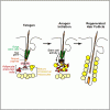
Yale researchers identified stem cells within the skin's fatty layer and showed that molecular signals from these cells were necessary to spur hair growth in mice. Men with male pattern baldness still have stem cells in follicle roots but these stem cells lose the ability to jump-start hair regeneration. Scientists have known that these follicle stem cells need signals from within the skin to grow hair, but the source of those signals has been unclear.

Researchers observed that when hair dies, the layer of fat in the scalp that comprises most of the skin's thickness shrinks. When hair growth begins, the fat layer expands in a process called adipogenesis. They found that a type of stem cell involved in creation of new fat cells -- adipose precursor cells -- was required for hair regeneration in mice. They also found these cells produce molecules called PDGF (platelet derived growth factors), which are necessary to produce hair growth.
MPB.gif


Festa E, Fretz J, Berry R, et al. Adipocyte Lineage Cells Contribute to the Skin Stem Cell Niche to Drive Hair Cycling. Cell 2011;146(5):761-71. ScienceDirect - Cell : Adipocyte Lineage Cells Contribute to the Skin Stem Cell Niche to Drive Hair Cycling

In mammalian skin, multiple types of resident cells are required to create a functional tissue and support tissue homeostasis and regeneration. The cells that compose the epithelial stem cell niche for skin homeostasis and regeneration are not well defined. Here, we identify adipose precursor cells within the skin and demonstrate that their dynamic regeneration parallels the activation of skin stem cells. Functional analysis of adipocyte lineage cells in mice with defects in adipogenesis and in transplantation experiments revealed that intradermal adipocyte lineage cells are necessary and sufficient to drive follicular stem cell activation. Furthermore, we implicate PDGF expression by immature adipocyte cells in the regulation of follicular stem cell activity. These data highlight adipogenic cells as skin niche cells that positively regulate skin stem cell activity, and suggest that adipocyte lineage cells may alter epithelial stem cell function clinically.
 
DeSantis AS, DiezRoux AV, Hajat A, et al. Associations of Salivary Cortisol Levels with Metabolic Syndrome and Its Components: the Multi-Ethnic Study of Atherosclerosis. Journal of Clinical Endocrinology & Metabolism. http://jcem.endojournals.org/content/early/2011/08/25/jc.2011-0483.abstract (Associations of Salivary Cortisol Levels with Metabolic Syndrome and Its Components: the Multi-Ethnic Study of Atherosclerosis)

Context: Prior research has identified associations between social-environmental factors and metabolic syndrome (MetS) components. The physiological mechanisms underlying these associations are not fully understood, but alterations in activity of the hypothalamic-pituitary-adrenal axis, a stress-responsive biological system, have been hypothesized to play a role.

Objective: The aim of the study was to determine whether MetS diagnosis and specific clusters of MetS components (waist circumference, high-density lipoproteins, glucose, and blood pressure) are associated with cortisol levels.

Design and Setting: We conducted cross-sectional analyses of data from the Multi-Ethnic Study of Atherosclerosis (MESA) study in the general community.

Patients or Other Participants: We studied a population-based sample of 726 adults (ages 48 to 89 yr) who do not have clinical diabetes.

Intervention(s): There were no interventions.

Main Outcome Measure(s): Cortisol awakening response, cortisol decline across the waking day, and total cortisol output were analyzed (using 18 timed measures of salivary cortisol over 3 d).

Results: Overall, we found little evidence that the presence of MetS or its components is related to cortisol output or patterns. Contrary to expectation, the presence of MetS was associated with lower rather than higher area under the curve, and no consistent pattern was observed when MetS components or subsets of components were examined in relation to cortisol.

Conclusions: Our findings do not support the hypothesis that differences in level or diurnal pattern of salivary cortisol output are associated with MetS among persons without clinical diabetes.
 
Turner L, Singh K, Garritty C, et al. An Evaluation of the Completeness of Safety Reporting in Reports of Complementary and Alternative Medicine Trials. BMC Complementary and Alternative Medicine 2011;11(1):67. http://www.biomedcentral.com/content/pdf/1472-6882-11-67.pdf

BACKGROUND: Adequate reporting of safety in publications of randomized controlled trials (RCTs) is a pre-requisite for accurate and comprehensive profile evaluation of conventional as well as complementary and alternative medicine (CAM) treatments. Clear and concise information on the definition, frequency, and severity of adverse events(AEs)is necessary for assessing the benefit-harm ratio of any intervention. Our objective, to assess the quality of safety reporting in CAM RCTs; to explore the influence of different trial characteristics on the quality of safety reporting.

METHODS: Survey of safety reporting in RCTs published in 2009 across 15 widely used CAM interventions identified from the Cochrane Collaboration's CAM Field specialized register of trials. The adequacy of reporting of AEs was defined and categorized according to the CONSORT for harms extension; the percentage of words devoted to the reporting of safety in the entire report and in the results section.

RESULTS: Two-hundred and five trials were included in the review. Of these, 15% (31/205) reported that no harms were observed during the trial period. Of the remaining 174 trials reporting any safety information, only 21% (36/174) had adequate safety reporting. For all trials, the median percentage of words devoted to the reporting of safety in the results section was 2.6. Moreover, 69% (n = 141) of all trials devoted a lesser or equal percentage of words to safety compared to author affiliations. Of the predictor variables used in regression analysis, multicenter trials had more words devoted to safety in the results section than single centre trials (P = 0.045).

CONCLUSIONS: An evaluation of safety reporting in the reports of CAM RCTs across 15 different CAM interventions demonstrated that the reporting of harms was largely inadequate. The quality of reporting safety information in primary reports of CAM randomized trials requires improvement.
 
Hypogonadotropic Hypogonadism in Type 2 Diabetes and Obesity

Subnormal free testosterone concentrations in association with inappropriately low Luteinizing Hormone (LH) and Follicle Stimulating Hormone (FSH) concentrations and a normal response to Gonadotropin Releasing Hormone (GnRH) of LH and FSH in type 2 diabetes were first described in 2004. These abnormalities were independent of the duration and severity of hyperglycemia [glycosylated hemoglobin (HbA1c)]. Magnetic resonance imaging in these hypogonadal patients showed no abnormality in brain or the pituitary. This association of hypogonadotropic hypogonadism (HH) with type 2 diabetes has now been confirmed in several studies and is present in 25–40% of these men. In this context, it is important that The Endocrine Society now recommends the measurement of testosterone in patients with type 2 diabetes on a routine basis. These observations were recently extended to younger patients with type 2 diabetes between the ages of 18 and 35 yr who had HH at a rate of 33% when the usual normal range for middle age was employed, whereas the rate was 58% when age-specific normal range for free testosterone for the young was employed. With the advent of more specific liquid chromatography tandem mass spectrometry assay for measuring total testosterone, the reference ranges for total and free testosterone have recently been revised downward. Using this methodology, in our most recent study, we have found that 29% of men with type 2 diabetes have subnormal free testosterone concentrations, as measured by equilibrium dialysis; 25% had HH, whereas only 4% had hypergonadotropic hypogonadism.

Type 2 diabetic men with low testosterone levels have also been found to have a high prevalence of symptoms suggestive of hypogonadism such as fatigability and erectile dysfunction. In all of the above studies, total testosterone and free testosterone concentrations were inversely related to body mass index (BMI) and age. However, the presence of low testosterone concentration was not entirely dependent upon obesity because 25% of nonobese patients (31% of lean and 21% of overweight) also had HH. HH is relatively rare in type 1 diabetes and, therefore, is not a function of diabetes or hyperglycemia per se. Thus, in view of the inverse relationship between BMI and testosterone concentrations in both type 1 and type 2 diabetes, HH is probably related to insulin resistance. Previous studies have shown that hypogonadism is associated with upper abdominal adiposity, insulin resistance, and the metabolic syndrome. Treatment of systemic insulin resistance by rosiglitazone leads to a modest increase in testosterone concentrations in men with type 2 diabetes, without the restoration of testosterone concentrations to normal.

A recent study investigated the prevalence of low testosterone concentrations in a large number of obese and diabetic men (mean age, 60 yr; range, 45–96 yr); 44% of diabetic and 33% of age-matched nondiabetic men had subnormal free testosterone concentrations, respectively. Forty percent of obese men and 50% of obese diabetic men had subnormal free testosterone concentrations. Thus, obesity is associated with a high prevalence of hypogonadism, and the presence of diabetes adds to that risk.

Low testosterone concentrations in men with type 2 diabetes are associated with an increased prevalence of symptoms of hypogonadism, obesity, very high CRP concentrations, mild anemia, and decreased BMD. In addition, these men have an elevated risk (two to three times) of cardiovascular events and death in two small studies. Short-term studies of testosterone therapy have demonstrated an increase in libido. In addition, there is an increase in insulin sensitivity. Some, but not all studies, have shown an improvement in glycemia, body composition, and cardiovascular risk factors such as cholesterol and CRP concentrations. Trials of a longer duration are clearly required to definitively establish the benefits and risks of testosterone replacement in patients with type 2 diabetes and HH.


Dandona P, Dhindsa S. Update: Hypogonadotropic Hypogonadism in Type 2 Diabetes and Obesity. Journal of Clinical Endocrinology & Metabolism 2011;96(9):2643-51. http://jcem.endojournals.org/content/96/9/2643.abstract

Studies over the last few years have clearly established that at least 25% of men with type 2 diabetes have subnormal free testosterone concentrations in association with inappropriately low LH and FSH concentrations. Another 4% have subnormal testosterone concentrations with elevated LH and FSH concentrations. The Endocrine Society, therefore, now recommends the measurement of testosterone in patients with type 2 diabetes on a routine basis. The subnormal testosterone concentrations are not related to glycosylated hemoglobin or duration of diabetes, but are associated with obesity, very high C-reactive protein concentrations, and mild anemia. In addition, subnormal testosterone concentrations in these men are associated with a two to three times elevated risk of cardiovascular events and death in two early studies. Short-term studies of testosterone therapy in hypogonadal men with type 2 diabetes have demonstrated an increase in insulin sensitivity and a decrease in waist circumference. However, the data on the effect of testosterone replacement on glycemic control and cardiovascular risk factors such as cholesterol and C-reactive protein concentrations are inconsistent. As far as sexual function is concerned, testosterone treatment increases libido but does not improve erectile dysfunction and thus, phosphodiesterase inhibitors may be required. Trials of a longer duration are clearly required to definitively establish the benefits and risks of testosterone replacement in patients with type 2 diabetes and low testosterone.
 
The Association Of Testosterone, Sleep, And Sexual Function

The machinery of reproduction is elaborate, involving a well orchestrated sequence of events. The biology of sexual function is an integral part of life satisfaction. In men and women, androgens appear to play an important role in sexual function and development. Testosterone, although defined as a male hormone, is essential to both sexes and is directly involved in several physiological effects, including the sense of well-being, motivation, muscle mass and strength, and cognition and memory.

As life expectancy has progressively increased at the turn of the 20th century for both men and women, it can be expected that more people will prolong their sexual activities. Thus, quality of life and general well-being are underlined by high expectations. However, it is not surprising that an increase in sexual complaints might also be accompanied by this substantial increase in lifespan. Indeed, impairment in sexual life due to age or medical illness may seriously hinder extended life satisfaction.

In addition to aging, the current global population works and lives under a 24/7 lifestyle that imposes less sleep and increases physical activities. Sleep deprivation has many consequences in humans, especially on the hormonal profile and possibly on sexual life. In addition, the search for sexual satisfaction has accelerated with widespread mainstream media coverage. The influence of testosterone on sexual behavior, mainly in males, has been documented for decades, but our knowledge about its interaction with sleep and the effects that sleep loss has on its concentrations is still limited. Moreover, the majority of hormone-associated sleep alterations occur mainly in women, because their reproductive lives include menstrual cycles, pregnancy and menopause transition, among other dynamic physiological conditions.

The main objective of the present review is to provide a comprehensive summary of the interaction between testosterone and sexual function, depicting how sleep loss and common lifestyle conditions of many societies can influence testosterone levels in both sexes and the effects on sexual behavior. Finally, they also review the literature of how this hormone is related to various health outcomes (e.g., erectile dysfunction (ED)) associated with sleep disturbances.


Andersen ML, Alvarenga TF, Mazaro-Costa R, Hachul HC, Tufik S. The association of testosterone, sleep, and sexual function in men and women. Brain Res. ScienceDirect - Brain Research : The association of testosterone, sleep, and sexual function in men and women

Testosterone has been the focus of several investigations and review studies in males, but few have addressed its effects on sleep and sexual function, despite evidence of its androgenic effects on circadian activity in both sexes. Studies have been conducted to understand how sleeping increases (and how waking decreases) testosterone levels and how this rhythm can be related to sexual function.

This review addresses the inter-relationships among testosterone, sexual function and sleep, including sleep-disordered breathing in both sexes, specifically its effects related to sleep deprivation. In addition, hormonal changes in testosterone that occur in the gonadal and adrenal axis with obstructive sleep apnea and other conditions of chronic sleep deprivation, and which consequently affect sexual life, have also been explored.

Nevertheless, hormone-associated sleep disruptions occur across a lifetime, particularly in women. The association between endogenous testosterone and sex, sleep and sleep disturbances is discussed, including the results of clinical trials as well as animal model studies. Evidence of possible pathophysiological mechanisms underlying this relationship is also described.

Unraveling the associations of sex steroid hormone concentrations with sleep and sexual function may have clinical implications, as sleep loss reduces testosterone levels in males, and low sex steroid hormone concentrations have been associated with sexual dysfunction.
 
Weight Watchers Works

Overweight patients in a Weight Watchers program succeeded in losing weight and improving disease risk factors such as blood lipids and insulin sensitivity relative to standard care in an international trial.

After one year, those randomized to the program lost an average of 5.06 kg (11.1 lbs), compared with an average loss of 2.25 kg (5.0 lb) with standard care, reported Susan A. Jebb, PhD, of the U.K. Medical Research Council's Human Nutrition Research division in Cambridge, England, and colleagues online in The Lancet.

Significant differences, all favoring the Weight Watchers group in the unblinded 772-patient trial, were also seen in levels of fasting insulin and glucose, and the ratio of total to HDL cholesterol.

Patients were referred and evaluated in primary care clinics in order to model real-world practice more closely than is often the case in trials of weight-reduction interventions.

"Data from our study suggest that referral of selected participants by a primary healthcare professional to a commercial weight loss programme that provides regular weighing, advice about diet and physical activity, motivation, and group support can offer a clinically useful early intervention for weight management in overweight and obese people that can be delivered at large scale," Jebb and colleagues wrote.

In an accompanying editorial, two other British researchers said the findings "are likely to be widely generalizable" since the Weight Watchers program is available in similar form in many countries, including the U.S.

"The study by Jebb and colleagues is the first to show that a commercial provider, for which people normally pay themselves, is more effective than primary care management, which is provided by taxation or health insurance," wrote Kate Jolly, MBChB, MSc, PhD, and Paul Aveyard, PhD, both of the University of Birmingham.

In the current study, the normal subscription fee was waived for participants assigned to Weight Watchers, but they paid for their own food -- unlike a previous trial of the Jenny Craig system in which meals were provided free of charge. http://www.medpagetoday.com/PrimaryCare/DietNutrition/22647

Jolly and Aveyard indicated that, given the study results, it would make sense for healthcare systems to pay the costs of such programs -- as is already the case in parts of Great Britain.

They also suggested that it may even be advisable for primary care providers to stop offering weight-loss management themselves -- although "this step would be premature in the absence of other evidence," they added.

The study took place in Great Britain, Australia, and Germany, with 377 individuals randomized to Weight Watchers and 395 to standard care. They were patients in primary care clinics, with their providers recommending them for weight-loss management either in the commercial program or to standard care from their usual providers.

The Weight Watchers program consisted of weekly community-based meetings in which a low-calorie balanced diet was promoted, along with exercise. Members selected their own goals for weight loss "with input from the group leader," Jebb and colleagues indicated.

Meetings included weigh-ins and group discussions to engender mutual support. Participants also had access to Internet-based tools for meal planning and monitoring caloric intake.

Standard care included diet and exercise counseling, based on national clinical guidelines, from primary care providers during clinic visits.

About 87% were women and participants' mean age was about 47. Average body mass index was 31, with mean fat mass of 33 kg (73 lb) and waist circumference of 100 cm (39.4 inches).

Outcome measures were reported primarily on the basis of intention-to-treat, with last observations carried forward for study dropouts.

Indeed, many patients in both groups failed to complete the study's full year. Some 39% of those assigned to Weight Watchers and 46% of those given standard care withdrew prematurely (P>0.05).

Those completing the study tended to show better results than the entire intention-to-treat sample. Weight loss among completers averaged 6.65 kg (14.6 lb) in the Weight Watchers group and 3.26 kg (7.2 lb) with standard care.

Conversely, the most rigorous analysis -- intention-to-treat with dropouts assumed to have lost no weight -- showed smaller losses in both groups: 4.06 kg (8.9 lb) with Weight Watchers and 1.77 kg (3.9 lb) with standard care.

Notably, though, the between-group differences in each case were similar and statistically significant (P<0.0001).

Jebb and colleagues also found in all three analyses that waist circumference and fat mass declined significantly.

Other biomarkers and risk factors improved more in the Weight Watchers group, although the specifics varied according to the analytical method.

With intention-to-treat and last observation carried forward, significantly greater benefits were seen with the commercial program for fasting insulin, fasting glucose, and the ratio of total to HDL cholesterol.

Weight Watchers also was a clear winner for insulin, LDL cholesterol, HDL cholesterol, and the ratio of total to HDL cholesterol -- but not for glucose -- both when analysis was restricted to participants completing the study and when it included all participants enrolled with dropouts assumed to have had no changes in these parameters from baseline.

In all three analyses, there were slight, statistically insignificant trends toward improvements in glycated hemoglobin, triglycerides, and blood pressure.

Limitations to the study included the allowable BMI range of 27 to 35, such that the results may not be generalizable to severely and morbidly obese people. Also, nearly 90% of participants were women, limiting the applicability to overweight men.

In addition, dropout rates varied substantially between countries: 25% among German participants versus 41% in Australia and 64% in Britain. Jebb and colleagues suggested that unique features in the British system may be have been responsible -- they cited "difficulties in scheduling of follow-up appointments in routine clinical practice," whereas follow-up in the other countries took place in specialist research centers.

Standard care also differed somewhat among the three countries, the researchers noted.

The study was funded by Weight Watchers International.


Jebb SA, Ahern AL, Olson AD, et al. Primary care referral to a commercial provider for weight loss treatment versus standard care: a randomised controlled trial. The Lancet. Primary care referral to a commercial provider for weight loss treatment versus standard care: a randomised controlled trial : The Lancet

Background - The increasing prevalence of overweight and obesity needs effective approaches for weight loss in primary care and community settings. We compared weight loss with standard treatment in primary care with that achieved after referral by the primary care team to a commercial provider in the community.

Methods - In this parallel group, non-blinded, randomised controlled trial, 772 overweight and obese adults were recruited by primary care practices in Australia, Germany, and the UK. Participants were randomly assigned with a computer-generated simple randomisation sequence to receive either 12 months of standard care as defined by national treatment guidelines, or 12 months of free membership to a commercial programme (Weight Watchers), and followed up for 12 months. The primary outcome was weight change over 12 months. Analysis was by intention to treat (last observation carried forward [LOCF] and baseline observation carried forward [BOCF]) and in the population who completed the 12-month assessment. This trial is registered, number ISRCTN85485463.

Findings - 377 participants were assigned to the commercial programme, of whom 230 (61%) completed the 12-month assessment; and 395 were assigned to standard care, of whom 214 (54%) completed the 12-month assessment. In all analyses, participants in the commercial programme group lost twice as much weight as did those in the standard care group. Mean weight change at 12 months was ?5•06 kg (SE 0•31) for those in the commercial programme versus ?2•25 kg (0•21) for those receiving standard care (adjusted difference ?2•77 kg, 95% CI ?3•50 to ?2•03) with LOCF; ?4•06 kg (0•31) versus ?1•77 kg (0•19; adjusted difference ?2•29 kg, ?2•99 to ?1•58) with BOCF; and ?6•65 kg (0•43) versus ?3•26 kg (0•33; adjusted difference ?3•16 kg, ?4•23 to ?2•11) for those who completed the 12-month assessment. Participants reported no adverse events related to trial participation.

Interpretation - Referral by a primary health-care professional to a commercial weight loss programme that provides regular weighing, advice about diet and physical activity, motivation, and group support can offer a clinically useful early intervention for weight management in overweight and obese people that can be delivered at large scale.


Rock CL, Flatt SW, Sherwood NE, Karanja N, Pakiz B, Thomson CA. Effect of a Free Prepared Meal and Incentivized Weight Loss Program on Weight Loss and Weight Loss Maintenance in Obese and Overweight Women. JAMA: The Journal of the American Medical Association 2010;304(16):1803-10. Effect of a Free Prepared Meal and Incentivized Weight Loss Program on Weight Loss and Weight Loss Maintenance in Obese and Overweight Women, October 27, 2010, Rock et al. 304 (16): 1803 — JAMA

Context The prevalence of overweight and obesity in the United States remains high. Commercial weight loss programs may contribute to efforts to reduce the prevalence of overweight and obesity, although few studies have examined their efficacy in controlled trials.

Objective To test whether a free prepared meal and incentivized structured weight loss program promotes greater weight loss and weight loss maintenance at 2 years compared with usual care.

Design, Setting, and Participants A randomized controlled trial of weight loss and weight loss maintenance in 442 overweight or obese women (body mass index, 25-40) aged 18 to 69 years (mean age, 44 years) conducted at US institutions over 2 years with follow-up between November 2007 and April 2010.

Intervention The program, which involves in-person center-based or telephone-based one-to-one weight loss counseling, was available over a 2-year period. Behavioral goals were an energy-reduced, nutritionally adequate diet, facilitated by the inclusion of prepackaged food items in a planned menu during the initial weight loss phase, and increased physical activity. Participants assigned to usual care received 2 individualized weight loss counseling sessions with a dietetics professional and monthly contacts.

Main Outcome Measures Weight loss and weight loss maintenance.

Results Weight data were available at 24 months for 407 women (92.1% of the study sample). In an intent-to-treat analysis with baseline value substitution, mean weight loss was 7.4 kg (95% confidence interval [CI], 6.1-8.7 kg) or 7.9% (95% CI, 6.5%-9.3%) of initial weight at 24 months for the center-based group, 6.2 kg (95% CI, 4.9-7.6 kg) or 6.8% (95% CI, 5.2%-8.4%) for the telephone-based group, and 2.0 kg (95% CI, 0.6-3.3 kg) or 2.1% (95% CI, 0.7%-3.5%) for the usual care control group after 24 months (P<.001 for intervention effect).

Conclusion Compared with usual care, this structured weight loss program resulted in greater weight loss over 2 years.


Wing RR. Treatment Options for Obesity. JAMA: The Journal of the American Medical Association 2010;304(16):1837-8. Treatment Options for Obesity, October 27, 2010, Wing 304 (16): 1837 — JAMA
 
Feero WG, Green ED. Genomics Education for Health Care Professionals in the 21st Century. JAMA: The Journal of the American Medical Association 2011;306(9):989-90. Genomics Education for Health Care Professionals in the 21st Century, September 7, 2011, Feero and Green 306 (9): 989 — JAMA

Recent genomic discoveries have brought about far-reaching advances in understanding the molecular basis of human health and disease. The vision for the future of genomics research developed by the National Human Genome Research Institute suggests more discoveries are likely to occur over the next few decades. 1 These insights have helped reveal remarkable and unexpected complexities of human biology; however, this scientific reality has slowed the immediate translation of genomic discoveries to health benefits. Nevertheless, the early implementation of genomic medicine has brought clinically important advances that rival any other period of discovery in the history of Western medicine. 2 For example, in 1990 the genetic and molecular basis was understood for fewer than 2% of the estimated 7000 suspected mendelian conditions; in 2011, approximately 40% of mendelian conditions have a known molecular basis.
 

Attachments

Men take note: If you want women to remember, speak to them in a low pitch voice. Then, depending on what they remember about you, they may or may not rate you as a potential mate. That's according to a new study by David Smith and colleagues from the University of Aberdeen in the UK. Their work shows for the first time that a low masculine voice is important for both mate choice and the accuracy of women's memory.

In a series of two experiments, Smith and colleagues show that memory in women is sensitive to male voice pitch, a cue important for mate choice because it can indicate genetic quality as well as signal behavioral traits undesirable in a long-term partner. These could include antisocial traits and lack of emotional warmth for example. In order to evaluate potential partners, women appear to rely on their memories to rapidly provide information about the attributes and past behavior of potential partners.

In the first experiment, 45 women were initially shown an image of a single object while listening to the name of the object spoken either by a high or low pitch male or female manipulated voice. They were then shown two similar but not identical versions of the object and asked to identify the one they had seen earlier. The women were also asked which voice they preferred.

In the second experiment, as well as manipulated voices, the researchers used real male and female voices to test how 46 new women rated the voices and how they scored on object memory.

In both cases, the authors found that women had a strong preference for the low pitch male voice and remembered objects more accurately when they have been introduced by the deep male voice.


Smith D, Jones B, Feinberg D, Allan K. A modulatory effect of male voice pitch on long-term memory in women: evidence of adaptation for mate choice? Memory & Cognition 2011:1-10. http://www.springerlink.com/content/j2t4m3882n1m6465/ (SpringerLink - Memory &amp; Cognition, Online First™)

From a functionalist perspective, human memory should be attuned to information of adaptive value for one’s survival and reproductive fitness. While evidence of sensitivity to survival-related information is growing, specific links between memory and information that could impact upon reproductive fitness have remained elusive. Here, in two experiments, we showed that memory in women is sensitive to male voice pitch, a sexually dimorphic cue important for mate choice because it not only serves as an indicator of genetic quality, but may also signal behavioural traits undesirable in a long-term partner. In Experiment 1, we report that women’s visual object memory is significantly enhanced when an object’s name is spoken during encoding in a masculinised (i.e., lower-pitch) versus feminised (i.e., higher-pitch) male voice, but that no analogous effect occurs when women listen to other women’s voices. Experiment 2 replicated this pattern of results, additionally showing that lowering and raising male voice pitch enhanced and impaired women’s memory, respectively, relative to a baseline (i.e., unmanipulated) voice condition. The modulatory effect of sexual dimorphism cues in the male voice may reveal a mate-choice adaptation within women’s memory, sculpted by evolution in response to the dilemma posed by the double-edged qualities of male masculinity.
 
Harmonization Of Guidelines For The Prevention And Treatment Of Cardiovascular Disease

The Canadian Cardiovascular Harmonized National Guideline Endeavour (C-CHANGE) Initiative harmonized and integrated more than 400 separate recommendations from 8 sets of guidelines into one comprehensive but simplified resource. Differing guidelines can lead to conflicting recommendations and become barriers to good treatment, and the guidelines group simplified and distilled practice and evidence down to 89 recommendations to help clinicians diagnose and manage risk factors for cardiovascular disease in their patients.

Harmonization and integration of guidelines is becoming increasingly important because of the growing emphasis on multidisciplinary care. Other regions and countries such as Europe, the United States and New Zealand are moving to harmonize guidelines.

The guidelines provide recommendations on screening, diagnostic strategies and treatment, including changes to health behaviours and pharmaceutical treatments for all the major cardiovascular risk factors that can lead to complications such as stroke, heart attack and sudden cardiac death.


Tobe SW, Stone JA, Brouwers M, et al. Harmonization of guidelines for the prevention and treatment of cardiovascular disease: the C-CHANGE Initiative. Canadian Medical Association Journal. http://www.cmaj.ca/content/early/2011/09/12/cmaj.101508.full.pdf

Cardiovascular disease is the most prevalent chronic medical condition in Canada, and evidence-based management of risk factors for cardiovascular disease can reduce morbidity and mortality. However, there are more than 400 individual recommendations for risk management of cardiovascular disease from various guidelines authored or sponsored by many different organizations. Because the guidelines were developed through multiple processes, they use different evidence grading systems, wording and emphasis. Thus, one can appreciate the challenge for health care providers who are managing the care of patients with, or at increased risk of, atherosclerotic diseases or with multiple comorbidities. Providers must determine which clinical practice guidelines to use, which risk factors to address first and which treatment targets to follow. This challenge created by multiple guidelines inhibits implementation, understanding and adherence, and hinders the delivery of clinically effective guideline-based care. Without a system in place for harmonization, there is a risk of conflicting recommendations for clinical practice. Even subtle differences in recommendations between guidelines have been identified as barriers to implementation.

For example, based on new evidence, acetylsalicylic acid is no longer routinely recommended for “patients with diabetes without coronary disease,” but it is still recommended in the guidelines of the Canadian Hypertension Education Program for “hypertensive patients without complications.” Meanwhile, the guideline for treatment of hypertension from the Canadian Hypertension Education Program recommends the use of a statin for all patients older than 40 years who have hypertension and three risk factors for cardiovascular disease; however, the guideline does not specify the actual treatment targets for low-density lipoprotein cholesterol. This is in contrast to the guidelines of the Canadian Diabetes Association and the Canadian Cardiovascular Society, which include treatment targets. Most of the guidelines include recommendations on exercise, but the recommendations are highly variable in the frequency, intensity, duration and types of exercises recommended.

The proliferation of conflicting, and often redundant, clinical practice guidelines is not unique to Canada. Although the problem has been recognized and potential processes to avoid these pitfalls have been suggested, single organizational approaches have had varied but limited success. In Canada, clinical practice guidelines have been developed by independent groups with interest in individual risk factors or diseases, usually working with or part of charitable nongovernment health organizations largely dependent on academic and community volunteers. Clinical practice guidelines often differ in terms of timing of screening, diagnostic approaches, treatment, and the details and descriptions of the foundational evidence that led to recommendations. Development of the guidelines often involves unnecessary duplication of work, such as appraisal of the literature, and exhausts scarce volunteer resources.
 
See! I told you so.

DeSantis AS, DiezRoux AV, Hajat A, et al. Associations of Salivary Cortisol Levels with Metabolic Syndrome and Its Components: the Multi-Ethnic Study of Atherosclerosis. Journal of Clinical Endocrinology & Metabolism. http://jcem.endojournals.org/content/early/2011/08/25/jc.2011-0483.abstract (Associations of Salivary Cortisol Levels with Metabolic Syndrome and Its Components: the Multi-Ethnic Study of Atherosclerosis)

Context: Prior research has identified associations between social-environmental factors and metabolic syndrome (MetS) components. The physiological mechanisms underlying these associations are not fully understood, but alterations in activity of the hypothalamic-pituitary-adrenal axis, a stress-responsive biological system, have been hypothesized to play a role.

Objective: The aim of the study was to determine whether MetS diagnosis and specific clusters of MetS components (waist circumference, high-density lipoproteins, glucose, and blood pressure) are associated with cortisol levels.

Design and Setting: We conducted cross-sectional analyses of data from the Multi-Ethnic Study of Atherosclerosis (MESA) study in the general community.

Patients or Other Participants: We studied a population-based sample of 726 adults (ages 48 to 89 yr) who do not have clinical diabetes.

Intervention(s): There were no interventions.

Main Outcome Measure(s): Cortisol awakening response, cortisol decline across the waking day, and total cortisol output were analyzed (using 18 timed measures of salivary cortisol over 3 d).

Results: Overall, we found little evidence that the presence of MetS or its components is related to cortisol output or patterns. Contrary to expectation, the presence of MetS was associated with lower rather than higher area under the curve, and no consistent pattern was observed when MetS components or subsets of components were examined in relation to cortisol.

Conclusions: Our findings do not support the hypothesis that differences in level or diurnal pattern of salivary cortisol output are associated with MetS among persons without clinical diabetes.
 
The Mislabelling Of Deoxycorticosterone - Making Sense Of Corticosteroid Structure And Function

Pick up any endocrinology text published in the last 60 years, and if it deals with the actions of the corticosteroids, you will usually read something to the effect that deoxycorticosterone (DOC, 11-deoxycorticosterone, cortexone, 21-hydroxyprogesterone) is a weak mineralocorticoid that has virtually no glucocorticoid activity. In fact, neither of these statements is true: DOC is both a potent mineralocorticoid and also a glucocorticoid. To the contrary, among the vertebrates and indeed some invertebrates as well, it may have an extraordinary array of functions. Such misleading views may have frustrated rational analysis of steroid hormone structure–function relationships for decades. Quite how this compound has been so misunderstood is an interesting puzzle that takes us back to the pioneer days of steroid hormone discovery.

DOC.gif

The structures of deoxycorticosterone (DOC), deoxycorticosterone acetate (DOCA), corticosterone and aldosterone (in two forms). According to a prevailing view, corticosterone is primarily a glucocorticoid, aldosterone primarily a mineralocorticoid, and DOC is a weak mineralocorticoid with no glucocorticoid activity. Since corticosterone differs from DOC only by the presence of an 11?-hydroxyl group, but aldosterone has a more complex acetal or hemiacetal structure, these functional designations defy rational interpretation of steroid structure–function relationships.


Vinson GP. The mislabelling of deoxycorticosterone: making sense of corticosteroid structure and function. Journal of Endocrinology 2011;211(1):3-16. The mislabelling of deoxycorticosterone: making sense of corticosteroid structure and function

Over the 70 or so years since their discovery, there has been continuous interest and activity in the field of corticosteroid functions. However, despite major advances in the characterisation of receptors and coregulators, in some ways we still lack clear insight into the mechanism of receptor activation, and, in particular, the relationship between steroid hormone structure and function remains obscure. Thus, why should deoxycorticosterone (DOC) reportedly be a weak mineralocorticoid, while the addition of an 11?-hydroxyl group produces glucocorticoid activity, yet further hydroxylation at C18 leads to the most potent mineralocorticoid, aldosterone? This review aims to show that the field has been confused by the misreading of the earlier literature and that DOC, far from being relatively inactive, in fact has a wide range of activities not shared by the other corticoids. In contrast to the accepted view, the presence of an 11?-hydroxyl group yields, in corticosterone or cortisol, hormones with more limited functions, and also more readily regulated, by 11?-hydroxysteroid dehydrogenase. This interpretation leads to a more systematic understanding of structure–function relationships in the corticosteroids and may assist more rational drug design.
 
Back
Top