OnLine First

Sainz J, Rudolph A, Hoffmeister M, et al. Effect of Type 2 Diabetes Predisposing Genetic Variants on Colorectal Cancer Risk. Journal of Clinical Endocrinology & Metabolism. http://jcem.endojournals.org/content/early/2012/03/09/jc.2011-2565.abstract (Effect of Type 2 Diabetes Predisposing Genetic Variants on Colorectal Cancer Risk)

Background: The link between colorectal cancer (CRC) and type 2 diabetes mellitus (T2D) has been extensively studied. Although it is commonly accepted that T2D is a risk factor for CRC, the underlying mechanisms are still poorly understood.

Research Design and Methods: Given that the genetic background contributes to both traits, it is conceivable that genetic variants associated with T2D may also influence the risk of CRC. We selected 26 T2D-related single-nucleotide polymorphisms (SNP) previously identified by genome-wide association studies and assessed their association with CRC and their interaction with known risk factors (gender, T2D, and body mass index) of CRC. Selected SNP were genotyped in 1798 CRC cases and 1810 controls from the population-based Darmkrebs: Chancen der Verhütung durch Screening (DACHS) study (Germany).

Results: Patients carrying the TCF7L2_rs7903146_T allele had an increased risk of CRC (Ptrend = 0.02), whereas patients harboring the IL13_rs20541_T allele had a reduced risk (Ptrend = 0.02). A further analysis revealed gender-specific effects: the TCF7L2_rs7903146_T allele was associated with an increased risk of CRC in women (Ptrend = 0.003) but not in men (Pinteraction = 0.06); theLTA_rs1041981_A allele was associated with a decreased risk for CRC in women (Ptrend = 0.02), with an opposite effect in men (Ptrend = 0.05; Pinteraction = 0.002); the CDKAL1_rs7754840_C allele was associated with a decreased risk for CRC in men (Ptrend = 0.03), with no effect in women (Pinteraction = 0.03). The risk associated with the presence of T2D was modified both by IGF2BP2_rs4402960 andPPAR?_rs1801282 SNP (Pinteraction = 0.04 and 0.04, respectively). None of the findings were significant after correction for multiple comparisons.

Conclusions: These findings suggest that T2D-related variants modify CRC risk independently and/or in an interactive manner according to the gender and the presence or absence of T2D.
 
Upchurch CT, Barrett EJ. Screening for Coronary Artery Disease in Type 2 Diabetes. Journal of Clinical Endocrinology & Metabolism. http://jcem.endojournals.org/content/early/2012/03/09/jc.2011-2122.abstract (Screening for Coronary Artery Disease in Type 2 Diabetes)

Clinical Context: Sensitive tools are available to diagnose occult ischemic and atherosclerotic coronary disease, yet screening for coronary artery disease (CAD) has not been shown to reduce cardiac events in patients with type 2 diabetes mellitus (T2DM). Professional guidelines are inconsistent regarding CAD screening recommendations, but it is suggested that those at highest risk (10-yr risk ?20%) for cardiac events may benefit.

Evidence Acquisition: We reviewed bibliographies of professional CAD screening guidelines, review articles, and clinical trials published within the last 10 yr, although we have included relevant older studies. We excluded studies that did not focus on T2DM or explicitly analyze that subgroup.

Evidence Synthesis: Although screening for coronary ischemia or atherosclerosis does provide incremental prognostic information in patients with T2DM and previously undiagnosed CAD, this has not been found to significantly impact outcomes. This appears to result from comparable efficacy of revascularization and optimal medical therapy in stable CAD. Limited evidence supports the hypothesis that those with more severe CAD (three-vessel, left main, proximal left anterior descending) amenable to bypass surgery may be potential beneficiaries of screening. However, the low prevalence of such candidates in the asymptomatic population, continuing advances with percutaneous intervention, and the lack of prospective trials makes such a recommendation currently unsupportable.

Conclusions: Findings to date do not support widespread screening for CAD in patients with T2DM. A future strategy identifying those at highest risk as screening candidates may ultimately be effective, but the optimal method for selecting those subjects or subsequent treatment is unknown.
 
Pivovarova O, Gogebakan O, Kloting N, et al. Insulin Up-Regulates Natriuretic Peptide Clearance Receptor Expression in the Subcutaneous Fat Depot in Obese Subjects: A Missing Link between CVD Risk and Obesity? Journal of Clinical Endocrinology & Metabolism.
http://jcem.endojournals.org/content/early/2012/03/09/jc.2011-2839.abstract (Insulin Up-Regulates Natriuretic Peptide Clearance Receptor Expression in the Subcutaneous Fat Depot in Obese Subjects: A Missing Link between CVD Risk and Obesity?)


Context: Natriuretic peptides (NP) regulate cardiovascular homeostasis and have multiple metabolic properties. Decreased levels of NP or “natriuretic handicap” are signs of insulin resistance such as central obesity. Increased expression of NP clearance receptor (NPRC) in sc adipose tissue (SAT) was observed in insulin-resistant subjects.

Objective: We hypothesized that insulin acutely regulates NP receptor expression in adipose tissue.

Design and Participants: NPRA, NPRB, and NPRC mRNA expression was measured in paired samples of visceral adipose tissue (VAT) and SAT from 157 subjects (108 with type 2 diabetes). The effect of insulin on NPR gene expression in SAT was studied in euglycemic-hyperinsulinemic and hyperglycemic-hyperinsulinemic clamp experiments. Additionally, the effect of insulin and glucose on NPR expression in the culture of primary human monocytes and macrophages was tested.

Results: NPRA and NPRC gene expression was higher in VAT compared with SAT (P < 0.01), but onlyNPRC gene expression strongly correlated with fasting insulin levels (r = 0.65, P = 0.04 × 10?3; and r = 0.54, P = 0.002, for VAT and SAT, respectively). NPRB expression was lower in VAT than in SAT in subjects with type 2 diabetes and was lower compared with nondiabetic subjects. NPRC gene expression was up-regulated in SAT during both euglycemic- and hyperglycemic-hyperinsulinemic clamps (P = 0.038 and P = 0.048, respectively), and was increased in high glucose and insulin treatment in monocytes (70.2%; P = 0.01), but not in mature macrophages.

Conclusion: Insulin increased expression of NPRC in SAT independently of circulating glucose concentrations. Thus, insulin might suppress circulating NP via up-regulation of NPRC expression in obesity, providing a novel link between hyperinsulinemia and obesity.
 
Burney BO, Hayes TG, Smiechowska J, et al. Low Testosterone Levels and Increased Inflammatory Markers in Patients with Cancer and Relationship with Cachexia. Journal of Clinical Endocrinology & Metabolism. http://jcem.endojournals.org/content/early/2012/03/09/jc.2011-2387.abstract (Low Testosterone Levels and Increased Inflammatory Markers in Patients with Cancer and Relationship with Cachexia)

Context: Male cancer patients suffer from fatigue, sexual dysfunction, and decreased functional performance and muscle mass. These symptoms are seen in men with hypogonadism and/or inflammatory conditions. However, the relative contribution of testosterone and inflammation to symptom burden in cancer has not been well-established.

Objective: The aim of this study was to measure testosterone levels in male cancer patients and determine the relationship between testosterone, inflammation, and symptom burden.

Design/Setting: This cross-sectional study enrolled patients from a tertiary-care center.

Subjects/Outcome Measures: Subjects included males with cancer-cachexia (CC; n = 45) and cancer without cachexia (CNC; n = 50), as well as noncancer controls (CO; n = 45). Total testosterone (TT), bioavailable testosterone, C-reactive protein (CRP), and IL-6 were measured in plasma. Functional performance was assessed by the ECOG (Eastern Cooperative Oncology Group) and KPS (Karnofsky Performance Scales), and sexual function was assessed by the IIEF (International Index of Erectile Function).

Results: Low testosterone levels were seen in more than 70% of CC cases. TT was lower in CC compared to CNC (P < 0.05). Also, CC had lower bioavailable testosterone, grip strength, IIEF scores, appendicular lean body mass, and fat mass and higher IL-6 and CRP compared to controls (P? 0.05). ECOG and KPS were lower in CC and CNC compared to controls (P ? 0.05). On multiple regression analysis, TT, albumin, and CRP predicted symptoms differentially in cancer patients.

Conclusions: CC patients have higher inflammation and lower testosterone, grip strength, functional status, erectile function, fat mass, and appendicular lean body mass. Inflammation, TT, and albumin are associated with heavier symptom burden in this population. Interventional trials are needed to determine whether testosterone replacement and/or antiinflammatory agents benefit cancer patients.
 
Cunningham RL, Lumia AR, McGinnis MY. Androgen Receptors, Sex Behaviour, and Aggression. Neuroendocrinology. http://content.karger.com/ProdukteDB/produkte.asp?Aktion=ShowPDF&ArtikelNr=337663&Ausgabe=0&ProduktNr=223855&filename=337663.pdf

Androgens are intricately involved in reproductive and aggressive behaviours, but the role of the androgen receptor in mediating these behaviours is less defined. Further, activity of the hypothalamic-pituitary-gonadal (HPG) axis and hypothalamic-pituitary-adrenal (HPA) axis can influence each other at the level of the androgen receptor. Knowledge of the mechanisms for androgens' effects on behaviours through the androgen receptor will guide future studies in elucidating male reproductive and aggressive behaviour repertoires.
 
Antioxidants As Inducers Of DNA Damage Response And Cell Death

Antioxidants found in fruits, vegetables and red wine killed cancer cells, including those that are resistant to treatment, in a study that scientists said may lead to more effective tumor fighters. Researchers from the U.S. National Institutes of Health identified 22 antioxidants that eradicated dividing cells, including two types that showed promise against drug-resistant cancer cells.


Fox JT, Sakamuru S, Huang R, et al. High-throughput genotoxicity assay identifies antioxidants as inducers of DNA damage response and cell death. Proceedings of the National Academy of Sciences. http://www.pnas.org/content/early/2012/03/12/1114278109.full.pdf

Human ATAD5 is a biomarker for identifying genotoxic compounds because ATAD5 protein levels increase posttranscriptionally in response to DNA damage. We screened over 4,000 compounds with a cell-based quantitative high-throughput ATAD5-luciferase assay detecting genotoxic compounds. We identified 22 antioxidants, including resveratrol, genistein, and baicalein, that are currently used or investigated for the treatment of cardiovascular disease, type 2 diabetes, osteopenia, osteoporosis, and chronic hepatitis, as well as for antiaging. Treatment of dividing cells with these compounds induced DNA damage and resulted in cell death. Despite their genotoxic effects, resveratrol, genistein, and baicalein did not cause mutagenesis, which is a major side effect of conventional anticancer drugs. Furthermore, resveratrol and genistein killed multidrug-resistant cancer cells. We therefore propose that resveratrol, genistein, and baicalein are attractive candidates for improved chemotherapeutic agents.
 
GH Replacement Therapy On Bone Mineral Density

GH deficiency (GHD) in hypopituitary adults results in multiple abnormalities in terms of body composition, bone mass, and glucose and lipid metabolism. GH replacement normalizes most of these abnormalities in adult populations including patients of various ages. The response to GH replacement may, however, vary in different subgroups of patients depending on the cause and severity of disease as well as on whether the disease was acquired in childhood or adulthood.

GH secretion declines with increasing age, but there are distinct differences between normal elderly subjects and elderly adults with structural hypothalamic–pituitary disease. The elderly GHD adults have lower GH secretion and increased total body fat compared with age-matched healthy subjects, whereas there is little difference in terms of lean mass. The results of several studies suggest that GH replacement in elderly GHD patients has approximately similar efficacy as that in younger GHD adults in terms of quality of life, body composition, and serum lipid pattern.

Young GHD adults have reduced bone mineral content (BMC) and bone mineral density (BMD). Short-term (?12 months) GH replacement in relatively young GHD adults results in unchanged or even decreased bone mass, whereas long-term GH replacement improves BMC and BMD in open studies. Meta-analyses, mainly of randomized GH treatment trials with relatively short duration, have shown either a moderate increase in lumbar spine BMD or no change in BMD, which could have been due to relatively small sample size. In elderly GHD adults not receiving GH replacement, bone mass and density are approximately similar to that in healthy age-matched controls. Little is known whether GH replacement affects BMC and BMD in elderly GHD adults. In a recent review of studies on elderly GHD adults, no significant effect of GH replacement on BMD was observed, but previous studies have been few and of short duration and/or included relatively few patients.

In this single-center, open-labeled, prospective study, the effect of 3-year GH replacement on bone mass and density was determined in 45 GHD patients >65 years at the beginning of study and in 45 matched younger control GHD patients. All patients had adult-onset GHD.


Elbornsson M, Gotherstrom G, Franco C, Bengtsson BA, Johannsson G, Svensson J. Effects of 3-year GH replacement therapy on bone mineral density in younger and elderly adults with adult-onset GH deficiency. Eur J Endocrinol 2012;166(2):181-9. http://eje-online.org/content/166/2/181.full

OBJECTIVE: Little is known of the effects of long-term GH replacement on bone mineral content (BMC) and bone mineral density (BMD) in elderly GH-deficient (GHD) adults.

DESIGN/PATIENTS/METHODS: In this prospective, single-center, open-label study, the effects of 3-year GH replacement were determined in 45 GHD patients >65 years and in 45 younger control GHD patients with a mean age of 39.5 (S.E.M. 1.1) years. All patients had adult-onset disease and both groups were comparable in terms of number of anterior pituitary hormonal deficiencies, gender, body mass index, and waist:hip ratio.

RESULTS: The mean maintenance dose of GH was 0.24 (0.02) mg/day in the elderly patients and 0.33 (0.02) mg/day in the younger GHD patients (P<0.01). The 3 years of GH replacement induced a marginal effect on total body BMC and BMD, whereas femur neck and lumbar (L2-L4) spine BMC and BMD increased in both the elderly and the younger patients. The treatment response in femur neck BMC was less marked in the elderly patients (P<0.05 vs younger group). However, this difference disappeared after correction for the lower dose of GH in the elderly patients using an analysis of covariance. There were no between-group differences in responsiveness in BMC or BMD at other skeletal locations.

CONCLUSIONS: This study shows that GH replacement increases lumbar (L2-L4) spine and femur neck BMD and BMC in younger as well as elderly GHD patients. This supports the notion that long-term GH replacement is also useful in elderly GHD patients.
 
Pennings B, Groen B, de Lange A, et al. Amino acid absorption and subsequent muscle protein accretion following graded intakes of whey protein in elderly men. American Journal of Physiology - Endocrinology And Metabolism 2012;302(8):E992-E9. http://ajpendo.physiology.org/content/302/8/E992.abstract (Amino acid absorption and subsequent muscle protein accretion following graded intakes of whey protein in elderly men)

Whey protein ingestion has been shown to effectively stimulate postprandial muscle protein accretion in older adults. However, the impact of the amount of whey protein ingested on protein digestion and absorption kinetics, whole body protein balance, and postprandial muscle protein accretion remains to be established. We aimed to fill this gap by including 33 healthy, older men (73 ± 2 yr) who were randomly assigned to ingest 10, 20, or 35 g of intrinsically L-[1-13C]phenylalanine-labeled whey protein (n = 11/treatment). Ingestion of labeled whey protein was combined with continuous intravenous L-[ring-2H5]phenylalanine and L-[ring-2H2]tyrosine infusion to assess the metabolic fate of whey protein-derived amino acids. Dietary protein digestion and absorption rapidly increased following ingestion of 10, 20, and 35 g whey protein, with the lowest and highest (peak) values observed following 10 and 35 g, respectively (P < 0.05). Whole body net protein balance was positive in all groups (19 ± 1, 37 ± 2, and 58 ± 2 ?mol/kg), with the lowest and highest values observed following ingestion of 10 and 35 g, respectively (P < 0.05). Postprandial muscle protein accretion, assessed by L-[1-13C]phenylalanine incorporation in muscle protein, was higher following ingestion of 35 g when compared with 10 (P< 0.01) or 20 (P < 0.05) g. We conclude that ingestion of 35 g whey protein results in greater amino acid absorption and subsequent stimulation of de novo muscle protein synthesis compared with the ingestion of 10 or 20 g whey protein in healthy, older men.
 
Butler MP, Karatsoreos IN, LeSauter J, Silver R. Dose-Dependent Effects of Androgens on the Circadian Timing System and Its Response to Light. Endocrinology. http://endo.endojournals.org/content/early/2012/03/28/en.2011-1842.abstract (Dose-Dependent Effects of Androgens on the Circadian Timing System and Its Response to Light)

The hypothalamic suprachiasmatic nucleus (SCN) is the locus of a master clock that regulates circadian rhythms in physiology and behavior. Gonadectomy in male mice lengthens the period of circadian rhythms and increases the day-to-day variability of activity onset time. Both of these responses are rescued by the nonaromatizable androgen dihydrotestosterone. Androgen receptors (AR) are localized in SCN neurons that receive direct retinal input. To explore how androgens affect circadian clock function and its responsiveness to photic cues, we measured wheel-running behavior and SCN AR expression in intact, gonadectomized, and testosterone-replaced mice, held under various photic conditions. Gonadectomy lengthened circadian period in constant dim light but not in constant darkness. Increasing intensities of constant light parametrically increased circadian period, and this was potentiated at all intensities by gonadectomy. In contrast, gonadectomy did not alter light-induced pupil constriction, suggesting a nonretinal locus of hormone action. In hormone-replaced animals housed in constant darkness, T concentration was positively correlated with precision of activity onset and with SCN AR expression and negatively correlated with duration of activity. We infer the existence of two androgenic mechanisms: one modulates SCN responsiveness to light, and the second modulates SCN timekeeping and locomotor activity in a dose-dependent manner. Finally, the effects of androgens on period are a result of hormonal modulation of the SCN's response to photic input rather than to a change in the inherent period of oscillators in the absence of light.
 
Cahill L. A Half-Truth Is a Whole Lie: On the Necessity of Investigating Sex Influences on the Brain. Endocrinology. http://endo.endojournals.org/content/early/2012/04/04/en.2011-2167.abstract (A Half-Truth Is a Whole Lie: On the Necessity of Investigating Sex Influences on the Brain)

Sex influences are proving to be extremely widespread on brain function, including the human brain. Ample evidence now proves that the sex of subjects can influence, ever reverse, findings, hence conclusions, at all levels of brain science, down to the molecular level, often in completely unanticipated ways. Thus the still-prominent assumption that sex influences may be safely ignored by neurobiologists is invalid and must be abandoned. The failure to properly consider the issue fills the literature with conclusions tenuous at best, false at worst. The continuing, widespread resistance to investigating sex influences among brain scientists, a resistance largely rooted in deeply entrenched biases against the topic, is becoming increasingly scientifically indefensible and strongly retards progress in our field.
 
Ikeda Y, Aihara K-i, Yoshida S, Akaike M, Matsumoto T. Effects of androgens on cardiovascular remodeling. Journal of Endocrinology. http://joe.endocrinology-journals.org/content/early/2012/04/05/JOE-12-0126.full.pdf

Androgens, the male sex hormones, exert various biological effects on many target organs through the transcriptional effects of the nuclear androgen receptor (AR). ARs are expressed not only in classical target organs, such as the brain, genital organs, bone, and skeletal muscles, but also in the cardiovascular system. Because the female sex hormones estrogens are well known to protect against cardiovascular disease, sex has been considered to have a significant clinical impact on cardiovascular mortality. However, the influence of androgens on the cardiovascular system has not been fully elucidated. To clarify this issue, we analyzed the effects of administration of angiotensin II and doxorubicin, an anticancer agent, in a loading model in male wild-type and AR-deficient mice. In this review, we focus on the actions of androgens as potential targets for the prevention of cardiovascular diseases in males.
 
This affirms a much earlier study - Long-term effects on testicular func... [J Clin Endocrinol Metab. 1996] - PubMed - NCBI

Hendriks AE, Laven JS, Boellaard WP, de Jong FH, Boot AM, Drop SL. [Fertility after high-dose sex steroid treatment to reduce final height]. Ned Tijdschr Geneeskd 2012;156(14):A3982. [Fertility after high-dose sex steroid... [Ned Tijdschr Geneeskd. 2012] - PubMed - NCBI

OBJECTIVE: To study the long term effects on fertility of high-dose sex steroid treatment to reduce final height.

METHODS: We compared 239 tall women and 60 tall men treated with high-dose sex steroids during puberty with respectively 174 untreated tall women and 56 untreated tall men. Reproductive history was assessed by interview in addition to ultrasound imaging of the testes or ovaries, endocrine parameters and semen analysis.

RESULTS: Treated women had a significantly lower chance of conception (odds ratio (OR): 0.22) and increased risk of fertility problems (OR: 2.29) and of receiving infertility treatment (OR: 3.44). In addition, treated women had a lower chance of having at least one live birth (OR: 0.26). Ovarian function was assessed in 174 tall women (119 treated). Thirty-nine women (23%) exhibited a hypergonadotropic profile. Treated women had a significantly greater chance of being diagnosed with primary ovarian insufficiency (OR: 2.83). These women had increased follicle stimulating hormone levels while antral follicle counts were decreased. The incidence of fertility problems in the treated men was low and comparable with untreated men. Testicular volume and sperm quality were comparable between the groups. Treated men, however, had reduced serum testosterone levels (13.3 vs. 15.2 nmol/l).

CONCLUSION: High-dose sex steroid treatment leads in the long term in women to an increased risk of fertility problems and involuntary childlessness. Treated women have a higher risk of exhibiting signs of primary ovarian insufficiency. In treated men fertility seems not to be affected, but their serum testosterone levels are significantly reduced.
 
Ghazi S, Zohdy W, Elkhiat Y, Shamloul R. Serum testosterone levels in diabetic men with and without erectile dysfunction. Andrologia. Serum testosterone levels in diabetic men with and without erectile dysfunction - Ghazi - 2012 - Andrologia - Wiley Online Library

Diabetes mellitus is a common chronic disease, affecting 0.5-2% worldwide. The Massachusetts Male Aging Study reported that up to 75% of men with diabetes have a lifetime risk of developing ED. Type 2 diabetes is associated with low total serum testosterone (TT) identified in several cross-sectional studies and systemic analyses.

There is a lack of consensus regarding what constitutes the lowest level of testosterone within the boundaries of normality. In this retrospective study, we sought to evaluate the effect of associated co-morbidities on serum total testosterone (TT) level in men with type 2 diabetes DM, either with or without erectile dysfunction (ED).

Three hundred and ninety-one patients were evaluated for erectile function using an abridged, five-item version of the International Index of Erectile Function-5. Measurements of TT, fasting lipid profile, blood sugar and glycated haemoglobin (HbA1c) were conducted. Penile hemodynamics was assessed using intracavernosal injection and penile duplex study.

Hypogonadism was found in 126 cases (33.2%), and normal TT was observed in 254 (66.8%). ED was detected in 119 cases in the hypogonadal group (94.4%) as compared to 155/254 (61.0%) in eugonadal group, P = 0.0001. TT was lower in diabetic men with ED as compared to those with normal erectile function (EF), 392.4 +/- 314.9 versus 524.3 +/- 140.2 ng dl(-1) , respectively, P < 0.0001.

After exclusion of patients with hypertension and dyslipidaemia, 185 men were evaluated, and there was no difference in the mean TT level among men with ED 490.6 +/- 498.2 ng dl(-1) versus normal EF 540.6 +/- 133.4 ng dl(-1) although, HbA1c remained lower in men with normal erectile function. Receiver operating characteristic (ROC) curve of TT in men without associated co-morbidities showed that EF was compromised at TT = 403.5 ng dl(-1) or less. Sensitivity of 63.3% and a specificity of 94.0% were detected. At this level, ED was found in 33/38 (86.8%) men with TT 403.5 ng dl(-1) , whereas ED was observed in 57/147 (38.8%) men with TT >/= 403.5 ng dl(-1) (P < 0.0001).

We propose a cut-off value of 403.5 ng dl(-1) of TT blood levels as an indicator for initiation of testosterone replacement therapy in diabetic men with ED. Further prospective controlled trials are recommended.
 
Last edited:
Role of Hydrogen Sulfide in the Physiology of Penile Erection

Hydrogen Sulfide (H2S) is implicated as the third neurotransmitter with a relaxant effect in cavernosal smooth muscle, suggesting the potential role of H2S in the physiology of erection. H2S synthesizing associated enzymes have been identified in penile tissue, confirming the L cysteine/H2S system in the physiology of erection. Unlike NO, main relaxant effect of H2S on smooth muscle is direct through activation of KATP and KCa channels. This provides an alternative approach for ED treatment, especially for those patients with poor or no response to PDE5i.

10120


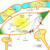
Distribution of H2S-producing associated enzymes in penile tissue H2S-producing associated enzymes were demonstrated to localize in smooth muscle cells (CBS, CSE) and nerve terminal (CSE) within the corpus caversnosum. H2S synthesizing associated enzymes including CBS, 3MTS and CAT were found in vascular endothelial cells. Even without direct evidence, endothelial cells in corpus cavernosum might contain these enzymes. Abbreviations: CBS= cystathionine ?-synthase, CSE= cystathionine ?-lyase, 3MTS=3- mercaptopyruvate sulfurtransferase, CAT= cysteine aminotransferase.

10121


Smooth muscle relaxant effect of H2S in corpus cavernosum. Three possible mechanisms are involved in H2S relaxing smooth muscle cells in corpus cavernosum: 1. Activating of KATP channel and KCa channel and inducing membrane hyperpolarization. 2. Inhibiting the activity of PDE5 and breakdown of cGMP. 3. Enhancing endothelial cells to release EDRFs or/and EDHFs. 4. Activation of TRPA1 ion channels. Abbreviations: PDE5= type 5 phosphodiesterase, cGMP=3’,5’-cyclic guanosine monophosphate, EDRFs= endothelium-derived relaxant factors, EDHFs= endothelium derived hyperpolarization factors, TRPA1= transient receptor potential A1.


Qiu X, Villalta J, Lin G, Lue TF. Role of Hydrogen Sulfide in the Physiology of Penile Erection. J Androl:jandrol.111.014936. Role of Hydrogen Sulfide in the Physiology of Penile Erection -- Qiu et al., 10.2164/jandrol.111.014936 -- Journal of Andrology

Hydrogen sulfide (H2S), which is a well known toxic gas, has recently been recognized as a biological messenger, which plays an important role in physiological and pathophysiological conditions. Relatively high levels of H2S have been discovered in mammalian tissues. It is mainly synthesized by two enzymes including cystathionine {beta}-synthase and cystathionine {gamma}-lysase, which utilize L-cysteine as substrate to produce H2S. H2S has been demonstrated to exhibit potent vasodilator activity both in vitro and in vivo by relaxing vascular smooth muscle. Recently, H2S has been discovered in penile tissue with smooth muscle relaxant effects. Furthermore, other effects of H2S may play a role in the physiology of erection. Understanding of H2S in the physiology of erection might provide alternative erectile dysfunction (ED) strategies for those patients with poor or no response to type 5 phosphodiesterase inhibitors (PDE5i). This review intends to present the H2S pathway in penile tissue and the potential role of H2S in the physiology of erections.
 

Attachments

  • H2S.gif
    H2S.gif
    89.5 KB · Views: 34
  • H2S-ED.gif
    H2S-ED.gif
    125.7 KB · Views: 34
Sartorius G, Spasevska S, Idan A, et al. Serum Testosterone, Dihydrotestosterone and Estradiol Concentrations in Older Men Self-Reporting Very Good Health:The Healthy Man Study. Clinical Endocrinology. Serum Testosterone, Dihydrotestosterone and Estradiol Concentrations in Older Men Self-Reporting Very Good Health:The Healthy Man Study - Sartorius - Clinical Endocrinology - Wiley Online Library

Objective To determine serum concentrations, intra-individual variability and impact of age-related co-morbidities on serum testosterone (T), dihydrotestosterone (DHT), estradiol (E2) and estrone (E1) in older men.

Design Observational, repeated measures study

Participants Men (n=325) 40 years and older self-reporting very good or excellent health.

Measurements Standardized history and physical examination, 9 blood samples at fixed time intervals over 3 months (three at 20 min intervals on days 1 (fasting) and 2 (non-fasting), one at days 7, 30 and 90). Serum T, DHT, E2 and E1 (n=2900, >99% of scheduled samples) measured by liquid chromatography-tandem mass spectrometry (LC-MS) were analyzed by linear mixed model analysis with fasting, age and obesity as covariables.

Results Mean serum T did not vary with age (p=0.76) but obesity (-0.35 nmol/L per body mass index (BMI) unit, p<0.0001) and ex-smoker status (-1.6 nmol/L, p<0.001) had significant effects. Serum DHT was increased with age (+0.011 nmol/L per year, p=0.001) but decreased with obesity (-0.05 nmol/L per BMI unit, p<0.0001). Serum E2 did not vary with age (p=0.31) or obesity (p=0.12). Overnight fasting increased (by 9-16%, all p<0.001) and reduced variability in morning serum T, DHT, E2 and E1. Non-fasting serum T and DHT were stable over time (day, week, month or 3 month; p>0.28).

Conclusions Serum T, DHT and E2 displayed no decrease associated with age among men over 40 years of age who self-report very good or excellent health although obesity and ex-smoking status were associated with decreased serum androgens (T, DHT) but not E2. These findings support the interpretation that the age-related decline in blood T accompanying non-specific symptoms in older men may be due accumulating age-related co-morbidities rather than a symptomatic androgen deficiency state.
 
Ya know, it would be a full time job just keeping up with all the excellent studies you post. :)
So much to do; so little time.
 
Reproductive Technologies and the Risk of Birth Defects

Consistent evidence from individual studies, including registry-based cohort studies and meta-analyses, has linked assisted conception involving in vitro fertilization (IVF) or intracytoplasmic sperm injection (ICSI) with an increased risk of birth defects. The associations between the use of these techniques and birth defects have appeared to be stronger for singleton births than for multiple births. It is unclear whether the excess of birth defects after IVF or ICSI may be attributable to patient characteristics related to infertility, rather than to the treatment, and whether the risk is similar across assisted reproductive technologies and related therapies. Available studies have been limited by small numbers of participants, the pooling of exposure groups, or, specifically for case–control studies, retrospective collection of data and questionable appropriateness of controls.

Investigators performed a population-wide cohort study, examining births and pregnancies terminated because of birth defects, to assess the risks of defects from pregnancy to a child's fifth birthday across a range of methods for treating infertility as compared with the risk associated with pregnancy that was not assisted by reproductive technology (spontaneous pregnancy). They also assessed the risks of birth defects associated with spontaneous pregnancy among women with a previous birth with assisted conception and women with a documented history of infertility but no treatment at assisted-reproductive-technology clinics.

In this large observational study using detailed Australian databases with information on several potential confounders, they confirmed previous findings of an increased risk of birth defects among births conceived with assisted reproductive technology as compared with births from spontaneous conception. After multivariate adjustment, the association between IVF and the risk of any birth defect was no longer significant, whereas the increased risk of any birth defect associated with ICSI remained significant.


Davies MJ, Moore VM, Willson KJ, et al. Reproductive Technologies and the Risk of Birth Defects. New England Journal of Medicine 2012;366(19):1803-13. MMS: Error

BACKGROUND - The extent to which birth defects after infertility treatment may be explained by underlying parental factors is uncertain.

METHODS - We linked a census of treatment with assisted reproductive technology in South Australia to a registry of births and terminations with a gestation period of at least 20 weeks or a birth weight of at least 400 g and registries of birth defects (including cerebral palsy and terminations for defects at any gestational period). We compared risks of birth defects (diagnosed before a child's fifth birthday) among pregnancies in women who received treatment with assisted reproductive technology, spontaneous pregnancies (i.e., without assisted conception) in women who had a previous birth with assisted conception, pregnancies in women with a record of infertility but no treatment with assisted reproductive technology, and pregnancies in women with no record of infertility.

RESULTS - Of the 308,974 births, 6163 resulted from assisted conception. The unadjusted odds ratio for any birth defect in pregnancies involving assisted conception (513 defects, 8.3%) as compared with pregnancies not involving assisted conception (17,546 defects, 5.8%) was 1.47 (95% confidence interval [CI], 1.33 to 1.62); the multivariate-adjusted odds ratio was 1.28 (95% CI, 1.16 to 1.41). The corresponding odds ratios with in vitro fertilization (IVF) (165 birth defects, 7.2%) were 1.26 (95% CI, 1.07 to 1.48) and 1.07 (95% CI, 0.90 to 1.26), and the odds ratios with intracytoplasmic sperm injection (ICSI) (139 defects, 9.9%) were 1.77 (95% CI, 1.47 to 2.12) and 1.57 (95% CI, 1.30 to 1.90). A history of infertility, either with or without assisted conception, was also significantly associated with birth defects.

CONCLUSIONS - The increased risk of birth defects associated with IVF was no longer significant after adjustment for parental factors. The risk of birth defects associated with ICSI remained increased after multivariate adjustment, although the possibility of residual confounding cannot be excluded.
 
Dai W, Li Y, Zheng H. Estradiol/Testosterone Imbalance: Impact on Coronary Heart Disease Risk Factors in Postmenopausal Women. Cardiology 2012;121(4):249-54. http://content.karger.com/produktedb/produkte.asp?doi=337274 (Estradiol/Testosterone Imbalance: Impact on Coronary Heart Disease Risk Factors in Postmenopausal Women)

Objective: Several groups have reported the important role of the estradiol/testosterone (E(2)/T) ratio in benign prostatic hyperplasia and cerebral vessels. However, there has been no study on the role of the E(2)/T ratio in women with coronary heart disease (CHD). This study aimed to evaluate the association among the ratio of sex hormones and known risk factors of atherosclerosis in postmenopausal women with CHD.

Methods: 114 controls and 124 postmenopausal women with CHD were selected for this study. Serum levels of estradiol, testosterone, aromatase, sex hormone-binding globulin (SHBG), lipid-lipoprotein profile and high-sensitivity C-reactive protein were determined.

Results: Compared with the control, the E(2)/T ratio decreased from 5.35 +/- 2.78 to 3.88 +/- 2.51 (p < 0.0001). Multiple linear regression analysis showed that the E(2)/T ratio was negatively associated with total cholesterol, low-density lipoprotein cholesterol (LDL-c) and the atherogenic index of plasma, but positively associated with high-density lipoprotein cholesterol (HDL-c) and HDL-c/LDL-c (for all, p < 0.0001). We found that there was a negative correlation between the E(2)/T ratio and aromatase (r = -0.192, p = 0.032) and a positive correlation between aromatase and SHBG (r = 0.938).

Conclusion: The balance of the serum E(2)/T ratio was broken in the women with CHD, and an imbalanced E(2)/T ratio showed a strong association with cardiovascular risk factors in postmenopausal women with CHD.
 
Song CH, Gong EY, Park JS, Lee K. Testicular Steroidogenesis is Locally Regulated by Androgen via Suppression of Nur77. Biochem Biophys Res Commun. ScienceDirect.com - Biochemical and Biophysical Research Communications - Testicular Steroidogenesis is Locally Regulated by Androgen via Suppression of Nur77

Steroidogenesis in the testis is regulated by a negative feedback mechanism through the hypothalamus-pituitary-testis axis. Recent studies suggest that besides this long-loop regulation, testicular steroidogenesis is also locally regulated by androgen. However, the molecular mechanism behind this additional regulatory pathway has been poorly addressed. In the present study, we demonstrate that liganded androgen receptor (AR) suppresses the transcriptional activity of Nur77 on steroidogenic enzyme gene promoters, affecting testicular steroidogenesis. AR physically interacts and colocalizes with Nur77 in the nucleus in the presence of androgen. AR inhibits Nur77 transactivation by competing mainly with coactivators such as SRC-1 for Nur77 binding. These results suggest that androgen, through binding to AR, directly acts as a signal inhibiting the expression of steroidogenic enzyme genes in Leydig cells, eventually resulting in decreased testicular steroidogenesis. These findings strongly support the hypothesis that androgen acts locally to regulate testicular steroidogenesis, and may provide its action mechanism.
 
Plasma HDL Cholesterol And Risk Of Myocardial Infarction

In summary, their results showed that polymorphisms related to plasma LDL cholesterol were consistently associated with risk of myocardial infarction, whereas this was not the case for variants related to plasma HDL cholesterol. A polymorphism in the endothelial lipase gene and a genetic score of 14 common SNPs that specifically raised HDL cholesterol were not associated with myocardial infarction, suggesting that some genetic mechanisms that raise HDL cholesterol do not lower risk of myocardial infarction. Hence, interventions (lifestyle or pharmacological) that raise plasma HDL cholesterol cannot be assumed ipso facto to lead to a corresponding benefit with respect to risk of myocardial infarction.


Voight BF, Peloso GM, Orho-Melander M, et al. Plasma HDL cholesterol and risk of myocardial infarction: a mendelian randomisation study. The Lancet. Plasma HDL cholesterol and risk of myocardial infarction: a mendelian randomisation study : The Lancet

Background - High plasma HDL cholesterol is associated with reduced risk of myocardial infarction, but whether this association is causal is unclear. Exploiting the fact that genotypes are randomly assigned at meiosis, are independent of non-genetic confounding, and are unmodified by disease processes, mendelian randomisation can be used to test the hypothesis that the association of a plasma biomarker with disease is causal.

Methods - We performed two mendelian randomisation analyses. First, we used as an instrument a single nucleotide polymorphism (SNP) in the endothelial lipase gene (LIPG Asn396Ser) and tested this SNP in 20 studies (20 913 myocardial infarction cases, 95 407 controls). Second, we used as an instrument a genetic score consisting of 14 common SNPs that exclusively associate with HDL cholesterol and tested this score in up to 12 482 cases of myocardial infarction and 41 331 controls. As a positive control, we also tested a genetic score of 13 common SNPs exclusively associated with LDL cholesterol.

Findings - Carriers of the LIPG 396Ser allele (2•6% frequency) had higher HDL cholesterol (0•14 mmol/L higher, p=8×10?13) but similar levels of other lipid and non-lipid risk factors for myocardial infarction compared with non-carriers. This difference in HDL cholesterol is expected to decrease risk of myocardial infarction by 13% (odds ratio [OR] 0•87, 95% CI 0•84—0•91). However, we noted that the 396Ser allele was not associated with risk of myocardial infarction (OR 0•99, 95% CI 0•88—1•11, p=0•85). From observational epidemiology, an increase of 1 SD in HDL cholesterol was associated with reduced risk of myocardial infarction (OR 0•62, 95% CI 0•58—0•66). However, a 1 SD increase in HDL cholesterol due to genetic score was not associated with risk of myocardial infarction (OR 0•93, 95% CI 0•68—1•26, p=0•63). For LDL cholesterol, the estimate from observational epidemiology (a 1 SD increase in LDL cholesterol associated with OR 1•54, 95% CI 1•45—1•63) was concordant with that from genetic score (OR 2•13, 95% CI 1•69—2•69, p=2×10?10).

Interpretation - Some genetic mechanisms that raise plasma HDL cholesterol do not seem to lower risk of myocardial infarction. These data challenge the concept that raising of plasma HDL cholesterol will uniformly translate into reductions in risk of myocardial infarction.
 
Back
Top