Anabolic Steroids & Liver [GI]

Herbal and Dietary Supplements-Induced Liver Injury

Background: Herbal and dietary supplements (HDS) consumption, a growing cause of hepatotoxicity, is a common practice among Latin-American populations.

Objectives: To evaluate clinical, laboratory features and outcome in HDS-hepatotoxicity included in the Latin America-Drug Induced Liver Injury (LATINDILI) Network.

Material and methods: A total of 29 adjudicated cases of HDS hepatotoxicity reported to the LATINDILI Network from October 2011 through December 2019 were compared with 322 DILI cases due to conventional drugs and 16 due to anabolic steroids as well as with other series of HDS-hepatotoxicity.

Results: From 367 DILI cases, 8% were attributed to HDS. An increasing trend in HDS-hepatotoxicity was noted over time (p=0.04). Camellia sinensis, Herbalife® products, and Garcinia cambogia, mostly used for weight loss, were the most frequently adjudicated causative agents. Mean age was 45 years (66% female). Median time to onset was 31 days.

Patients presented typically with hepatocellular injury (83%) and jaundice (66%). Five cases (17%) developed acute liver failure. Compared to conventional medications and anabolic steroids, HDS hepatotoxicity cases had the highest levels of aspartate and alanine transaminase (p=0.008 and p=0.021, respectively), had more re-exposure events to the culprit HDS (14% vs 3% vs 0%; p=0.026), and had more severe and fatal/liver transplantation outcome (21% vs 12% vs 13%; p=0.005). Compared to other DILI cohorts, less HDS hepatotoxicity cases in Latin America were hospitalized (41%).

Conclusions: HDS-hepatotoxicity in Latin-America affects mainly young women, manifests mostly with hepatocellular injury and is associated with higher frequency of accidental re-exposure. HDS hepatotoxicity is more serious with a higher chance of death/liver-transplantation than DILI related to conventional drugs.

Bessone F, García-Cortés M, Medina-Caliz I, et al. HERBAL AND DIETARY SUPPLEMENTS-INDUCED LIVER INJURY IN LATIN AMERICA: EXPERIENCE FROM THE LATINDILI NETWORK. Clin Gastroenterol Hepatol. 2021 Jan 9:S1542-3565(21)00013-6. doi: 10.1016/j.cgh.2021.01.011. Epub ahead of print. PMID: 33434654. https://www.cghjournal.org/article/S1542-3565(21)00013-6/pdf
 
[OA] Chemical Risk Factors of Primary Liver Cancer

Primary liver cancer has the sixth highest incidence and fourth highest cancer mortality worldwide. Hepatitis B is the leading cause of liver cancer, though its incidence is decreasing with vaccination.

Alcohol is the leading cause of liver transplant, cirrhosis, and cancer in the developed world, and is projected to surpass hepatitis B as the leading hepatic cancer etiology worldwide. Tobacco smoking has shown a positive association with liver cancer in a majority of studies, though not all.

Aflatoxin, a mycotoxin produced by Aspergillus, is estimated to account for 3-20% of global liver cancer cases, 40% of which occur in sub-Saharan Africa. These statistics are confounded by the prevalence of hepatitis B, which may have a synergistic effect on hepatic carcinogenesis. Aflatoxin is ingested and likely inhaled from agricultural products, placing farmers, food processors, and textile workers in developing nations at risk.

Vinyl-chloride is used in the production of PVC plastics and causes rare liver angiosarcoma, hepatocellular carcinoma, and other neoplasms. Arsenic and cadmium are naturally-occurring, hepatocarcinogenic metals with high occupational exposure in industries involving coal, metals, plastics, and batteries.

Millions of laborers in waste-disposal and manufacturing are exposed to organic solvents and N-nitrosamines, which vary from carcinogenic (group 1) to possibly carcinogenic (group 2B) in their IARC designation. Insecticide DDT is possibly hepatocarcinogenic (group 2B), though continues to be used for malaria control in the developing world.

While suggested by case reports, anabolic steroids and oral contraceptives have not been shown to increase liver cancer risk in large studies.

Barsouk A, Thandra KC, Saginala K, Rawla P, Barsouk A. Chemical Risk Factors of Primary Liver Cancer: An Update. Hepat Med. 2021 Jan 5;12:179-188. doi: 10.2147/HMER.S278070. PMID: 33447099; PMCID: PMC7801911.
https://www.dovepress.com/chemical-risk-factors-of-primary-liver-cancer-an-update-peer-reviewed-fulltext-article-HMER
 
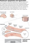
For organ homeostasis or regrowth after injury or disease, one or more stem cell populations is needed to rebuild lost tissue. There is considerable debate about the source of new cells in the liver. Two groups now identify the source of new hepatocytes. Although the liver may seem to lack major variation across its structure, its lobule is organized into concentric zones where hepatocytes express different metabolic enzymes.

Wei et al. sought to systematically define the source of new liver cells by comparing 14 fate-mapping mice that label different liver cell types. They found that different regions of the liver lobule exhibit differences in hepatocyte turnover, with zone 2 representing a primary source of new hepatocytes during homeostasis and regeneration.

Similarly, He et al. designed a genetic approach to record cell proliferation in vivo with high spatial and temporal resolution to enable continuous recording of proliferative events of any specific cell type at the whole-cell population level. Using this method, they identified zone 2 as having the highest proliferative activity and contributing the most to liver regrowth.

These findings have implications for the cellular basis of chronic disease pathogenesis, cancer development, and regenerative medicine strategies.


In The Zone For Liver Proliferation

In humans, the liver is the most regenerative solid organ, able to regrow to normal size after removal of up to 90% of the liver volume. The liver is also distinct because it scales with body size, a characteristic that has been attributed to a “hepatostat” that adjusts liver size to the needs of the body. Identifying the cells contributing to liver homeostasis and repair could lead to therapies that can activate specific cellular compartments responsible for regeneration.

Wei et al. (Liver homeostasis is maintained by midlobular zone 2 hepatocytes | Science) and He et al. (Proliferation tracing reveals regional hepatocyte generation in liver homeostasis and repair | Science), respectively, find that, in mice, a subset of cells in a particular region of the liver, called midlobular zone 2, are the major contributors to hepatocyte proliferation during homeostasis and identify other hepatocyte subsets that contribute to regeneration after damage. This raises questions regarding the mechanisms that induce hepatocyte proliferation and the zonal division of labor with respect to the hepatostat.

Andersson ER. In the zone for liver proliferation. Science 2021;371:887. In the zone for liver proliferation | Science

F1.large.jpg
 

Attachments

Proliferation Tracing Reveals Regional Hepatocyte Generation in Liver Homeostasis and Repair

Organ homeostasis is orchestrated by time- and spatially restricted cell proliferation. Studies identifying cells with superior proliferative capacities often rely on the lineage tracing of a subset of cell populations, which introduces a potential selective bias.

In this work, we developed a genetic system [proliferation tracer (ProTracer)] by incorporating dual recombinases to seamlessly record the proliferation events of entire cell populations over time in multiple organs. In the mouse liver, ProTracer revealed more hepatocyte proliferation in distinct zones during liver homeostasis, injury repair, and regrowth.

Clonal analysis showed that most of the hepatocytes labeled by ProTracer had undergone cell division. By genetically recording proliferation events of entire cell populations, ProTracer enables the unbiased detection of specific cellular compartments with enhanced regenerative capacities.

He L, Pu W, Liu X, et al. Proliferation tracing reveals regional hepatocyte generation in liver homeostasis and repair. Science 2021;371:eabc4346. Proliferation tracing reveals regional hepatocyte generation in liver homeostasis and repair | Science


Liver Homeostasis Is Maintained by Midlobular Zone 2 Hepatocytes


The liver is organized into zones in which hepatocytes express different metabolic enzymes. The cells most responsible for liver repopulation and regeneration remain undefined, because fate mapping has only been performed on a few hepatocyte subsets.

Here, 14 murine fate-mapping strains were used to systematically compare distinct subsets of hepatocytes. During homeostasis, cells from both periportal zone 1 and pericentral zone 3 contracted in number, whereas cells from midlobular zone 2 expanded in number. Cells within zone 2, which are sheltered from common injuries, also contributed to regeneration after pericentral and periportal injuries.

Repopulation from zone 2 was driven by the insulin-like growth factor binding protein 2–mechanistic target of rapamycin–cyclin D1 (IGFBP2-mTOR-CCND1) axis. Therefore, different regions of the lobule exhibit differences in their contribution to hepatocyte turnover, and zone 2 is an important source of new hepatocytes during homeostasis and regeneration.

Wei Y, Wang YG, Jia Y, et al. Liver homeostasis is maintained by midlobular zone 2 hepatocytes. Science 2021;371:eabb1625. Liver homeostasis is maintained by midlobular zone 2 hepatocytes | Science
Dear doctor, can you please help me with finding full text of one study?
 
What is the opinion on 5% Nutrition Liver & Organ defender? Maybe better to just stick with the tried and true TUDCA, NAC and Milk Thistle.
 
So obviously tudca and NAC have some real studies and benefits behind them but it begs the question, if it is as beneficial as they say why is it not a prescription medication? Wouldn’t the medical industry try to gatekeep it from being so accessible? Or do they just have much better more effective prescription medications than the latter ?
 
After reading this thread you made think twice about running dbol in my upcoming cycle. I am getting up there in age so I was only going to run it in my first 3-4 weeks to kick start things but I may have to do without ... Or atleast shorten the duration of usage. Very good read ... Thanks for sharing
I agree just not worth it. I personally am lot more patient for just waiting for the injectables to get in the blood rather than take a kick start
 
Back
Top